Download
1 / 29
300 likes | 400 Views
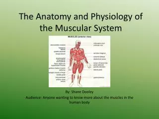
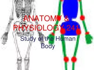
Anatomy & physiology. Muscles : Types and Fibres. Muscles. There is three types of muscle; Cardiac- in-voluntary the muscle that encompass the heart Smooth- in-voluntary Skeletal- voluntary Provides Human Movement, attached to the skeleton by tendons.

E N D
Anatomy & physiology Muscles: Types and Fibres
Muscles • There is three types of muscle; • Cardiac- in-voluntary the muscle that encompass the heart • Smooth- in-voluntary • Skeletal- voluntary Provides Human Movement, attached to the skeleton by tendons Divide the class into 3 (Cardiac/Smooth and Skeletal muscle): - Find a couple of images of the muscle - Say what this type of muscle does - Give a few examples Be prepared to feedback your findings to the class…
Cardiac Muscle The heart has its own muscle tissue called Cardiac/Myocardium muscle. It is an Involuntary muscle: as it has its own nerve supply. It works by sending nerve impulses through the cells and fibres within the muscle. Function: the function of the myocardium is to pump blood around the body. It beats 60-80 times a minute
Also an involuntary muscle as they are out of out CONSCIOUS control. Smooth Muscle Found in the digestive system (large and small intestine), Circulatory system (artery and vein walls) and Urinary system. Smooth muscle contracts consecutively (peristaltic action), producing a wavelike effect. E.G. when food is passed through the digestive system it is slowly squeezed through the intestines.
Skeletal Muscle- Muscle that is attached to the skeleton across joints… It is under voluntary control as we decide when to contract the muscles and produce movement. Co-ordinated contractions of the skeletal muscle allow us to move smoothly and produce sports skills. • Skeletal muscle is responsible for the following functions: • Produce movement • Maintaining body posture • Generate heat for warmth • Storage of Glycogen for ENERGY Over 700 skeletal muscles make up 40% of our body weight (slightly less for females).
Types of Skeletal Muscle: • Within our skeletal muscle we actually have 2 types of muscle fibre called fast and slow twitch fibres, related to the speed in which they contract. Slow Fibres: Walk long distances Fast fibres: Chase our prey when needed Both these fibre types go back to when we were HUNTERS and GATHERERS
Types of Skeletal muscle- Task… You will use all of the different types of muscle fibres during a game of football. However, some fibres are particularly associated with different sports. Type I fibres are typically employed in a warm up and at the beginning of exercise. Type IIa fibres can be used for moderate intensity exercise. They are also often used for low intensity aerobic activities when we are very fatigued and the Type I fibres are tired. Type IIb fibres are used in explosive, powerful, fast movements. They tire very quickly. In the space below give 5 sports that are primarily associated with each muscle fibre type. Then give at least 2 examples of a footballer using each type of muscle fibre on match day.
Type 1- Slow Twitch Fibres RED in colour, as they have a good blood supply They are suited to endurance work and are slow to fatigue- Due to having a dense network of blood vessels. They also contain many MITOCHONDRIA(Energy producing organelles within cells), making them more efficient at producing energy using OXYGEN (O2).
Type 2a and type 2b- Fast twitch Fibres They have a poor blood supply, meaning they are whiter in appearance and will fatigue quicker due to lack of OXYGEN (O2) • Fast twitch fibres contract twice as quickly as slow twitch fibres and THICKER in size. Their FASTER, HARDER contractions make them suitable for producing fast and powerful contractions. E.G: Sprinting and Weightlifting
Type 2 (b) Type 2 (a) These fibres work when a person is working close to their maximum intensity. For example a 100m runner would use these type of fibres, or an Olympic lifter performing a fast lift. Work at slightly lower intensities, but higher than slow twitch fibres are capable of. For example a 400m runner would utilise Type 2A fibres.
Training effects for muscle fibres… Type 1 and Type 2b fibres will always retain their distinctive features… However Type 2a can take on characteristics of Type 1 and Type 2b depending on the training done (they do not change their fibre type). Postural muscles (muscles that keep us standing upright) like the muscles in the legs, back and abdominal areas will be predominantly SLOW TWITCH. As they produce low forces over a long period of time. The type of muscles found in the legs will determine whether you are more suited to sprinting or endurance running. Your athletic performances will be a good indicator of which. Bursztyn (1997): well trained middle- distance athletes will have 80% slow twitch fibres and well trained sprinters may have up to 75% fast twitch fibres
Anatomy and Physiology • Muscles: Movement
Understanding muscle action Muscles are attached to bones by tendons. The tendon at the non-moving (or fixed) end is known as the origin. The tendon at the moving end is known as the insertion. Muscles pull by contracting – they cannot push to produce the opposite movement. Muscles are arranged in antagonistic pairs. As one muscle contracts (shortens) its partner relaxes (lengthens). They swap actions to reverse the movement.
Muscular Movement • To cause movement, muscles must work across the joint. • For example, the bicep works across the elbow joint causing flexion of the elbow (bending) • When the muscle contracts it pulls on the bone, causing movement. • The bones act like levers, and the joints are the fulcrum. • The strength of the contraction depends on the amount of muscle fibres brought into use. • This is known as muscular fibre recruitment.
Upper arm muscles The biceps and triceps work together as an antagonistic pair to move the elbow joint. To flex the elbow, the biceps (the flexor) contracts and the triceps (the extensor) relaxes. To extend the elbow, the actions are reversed so that the triceps contracts and the biceps relaxes.
Quadriceps / hamstring muscle action The quadriceps and hamstrings in the legs are another antagonistic pair. Can you answer the following questions? Which joint do they move? quadriceps What types of movement are produced? hamstrings Which is the flexor and which is the extensor? Identify the origin and insertion of each muscle.
Antagonistic Action • Each muscle involved in the movement of a specific body part has a role: • Agonist/Prime Mover Muscle: this is the muscle that courses the movement e.g the bicep causes the movement during a bicep curl • Antagonist/Opposing Muscle: the opposing muscle must counter the action of the primer mover muscle to allow the action to take place e.g. the triceps must relax to allow for the biceps to contract during a bicep curl. • Fixator Muscle: this muscle works to stabilise the joint at the origin of the prime mover muscle e.g. the trapezius contracts to stabilise the origin of the biceps during the bicep curl. • Synergists:this muscle helps the prime mover to produce the desired movement by preventing any undesirable movements., during the upward phase of the bicep curl the brachialis muscle is the synergist
Types of contractions • There are 4 main types of contraction 1. Concentric Contraction: Most common contraction, it takes place when the ends of the muscle come closer together and the muscle shortens 2. Eccentric Contraction: The muscle ends move further away from each other 3. Isometric Contractions: A muscle exerts a force but does not change in length E.G.during a tug of war everyone is pulling the rope and your arm muscles are contracting to do this but your muscles are not shortening or lengthening. A rugby league example would be when both sides push in a scrum.
Isotonic Contractions: Eccentric and Concentric contractions Video of an eccentric, concentric and isometric contraction Concentric- Contraction to push weight away from the body Eccentric- Involves controlling the weight on it’s way down
Isometric contraction • Where a muscle contracts, but does not change in length • The muscle is active in holding a static position • This is easy to train, but soon leads to fatigue
P4 To achieve M1 you need to explain the function of the muscular system and the three different fibre types by giving reasons to support your description of how the systems work To achieve P4 you need to describe: • The different types of muscle (i.e. cardiac, skeletal, smooth). • The different types of muscle fibre (i.e. Type 1, Type2 and Type 2a) and the different types of sporting activity that would use those fibre types. • The roles that muscles can adopt during movement and the different contraction types. To achieve D1 you need to analyse the function of the muscular system and the three different fibre types by identifying all the factors and saying how they are related to one another. Describe what contribution they make to the system and give practical examples to support your argument.
Muscles • Muscles underpin human movement in all manners from picking up a pen to striking a ball. • An athletes ability to move muscle efficiently and effectively in unison can often be the difference between winning and loosing http://www.youtube.com/watch?v=Z-xesd1er1s 7.10
Muscular system • Research task: Individually research the chosen muscle. Locate it on the human body and talk about what it does in relation to the muscular system. You will then be asked to draw it on the whiteboard at the front of the class and explain your findings biceps triceps deltoids pectorals rectus abdominus quadriceps. These must be split into: o rectus femoris o vastuslateralis o vastusmedialis o vastusintermedius) hamstrings. These must split into: · semimembranosus · semitendinosus · biceps femoris gastrocnemius soleus tibialis anterior erector spinae teres major trapezius latissimusdorsi obliques gluteus maximus Use picture of body in paint…
MUscular system • Lower body: • Anterior view • Quadriceps: • Rectus Femoris • vastusIntermedius • VastusLateralis • VastusMedialis Tibialis anterior
Muscular System • Posterior View (back) • Lower body Gluteus Maximus • Hamstrings: • Semimembranosus • Semitendinosus • Biceps Femoris Gastrocnemius Soleus
Muscular System • Upper body anterior view Biceps Triceps Deltoids Pectoralis Major Rectus Abdominus Obliques
Muscular System • Upper body posterior view Teres Major Trapezius- Upper back LatisimusDorsi- Lower Back Erector spinae
Task… To achieve P3 you need to identify: • The location of the major muscles (biceps, triceps, deltoids, pectoralis major, rectus abdominis, rectus femoris, vastuslateralis, vastusmedialis, vastusintermedius, semimembranosus, semitendinosus, biceps femoris, gastrocnemius, soleus, tibialis anterior, erector spinae, teres major, trapezius, latissimusdorsi, obliques, gluteus maximus) in the body (label a diagram)