Download
1 / 17
170 likes | 178 Views
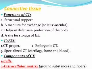
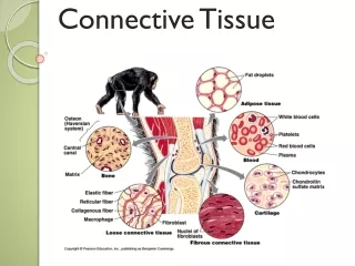
Lab 3 Connective tissue (2). Cartilage,Bone and Blood smear. Jun Zhou( 周俊) , Ph.D & M.D School of Medicine,Zhejiang University. Slides. 1.cartilage ( No.3 trachea) 2.Bone (No.5) 3.Blood smear (picture drawing). Hyaline Cartilage. C-shape hyaline cartilage. Trachea (No.3).

E N D
Lab 3 Connective tissue (2) Cartilage,Bone and Blood smear Jun Zhou(周俊), Ph.D & M.D School of Medicine,Zhejiang University
Slides • 1.cartilage ( No.3 trachea) • 2.Bone (No.5) • 3.Blood smear (picture drawing)
Hyaline Cartilage C-shape hyaline cartilage Trachea (No.3)
Hyaline Cartilage Trachea(No.3):H&E ×40
capsule lacuna perichondrium Isogenous groups Trachea:Hyaline Cartilage H&E ×400
Bone • Haversian system(osteon) • Central canal • Circumferential lamella • Interstitial lamella • Bone lacuna • Bone canaliculus Silver ×100
Haversian system(osteon) • Central canal • Bone lacuna • Bone canaliculus Bone:Silver ×400
perforating (Volkmann’s) cannals Bone:Silver ×100
Blood smear: A drop of blood is placed directly on a glass slide and spread over its surface with the edge of another slide. Stained with Wright’s stain.
Erythrocytes: • 7-8um • Biconcave disc • Stain uniformly with eosin • Neutrophil: • proportion - 50-70%; • Most numerous • Multilobed nucleus • Pale pink granules Blood smear:Wright ×400
Eosinophils. • 2-4% • 8-10 microns morphology • bilobed nucleus large, • uniform eosinophil granules. Blood smear:Wright ×400
Basophils. • Proportion 0.5% • morphology - large, irregular basophil granules obscure the nucleus Blood smear:Basophil Giemsa stain ×400
Lymphocyte: • Intensely stained nucleus • Possess a slight indentation • Small amount of cytoplasm • Pale blue Blood smear:Wright ×400
Monocyte: • nucleus ovoid to kidney shaped • blue-gray Cytoplasm Blood smear:Wright ×400
Platelets: • Small speck-like objectives • Aggregated into small groups • From megakaryocytes in myeloid tissue. Blood smear:Wright ×400