Download
1 / 1
10 likes | 74 Views
Results. Mechanical Properties of the Developing Porcine Skull Arun Mahapatra, Tim Baumer, B.S., Dr. Roger Haut, Ph.D.

E N D
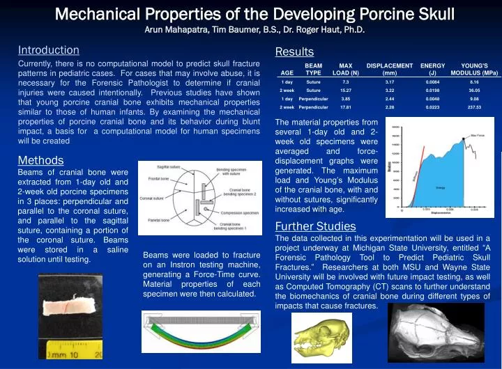
Results Mechanical Properties of the Developing Porcine SkullArun Mahapatra, Tim Baumer, B.S., Dr. Roger Haut, Ph.D. The material properties from several 1-day old and 2-week old specimens were averaged and force-displacement graphs were generated. The maximum load and Young’s Modulus of the cranial bone, with and without sutures, significantly increased with age. Methods Beams of cranial bone were extracted from 1-day old and 2-week old porcine specimens in 3 places: perpendicular and parallel to the coronal suture, and parallel to the sagittal suture, containing a portion of the coronal suture. Beams were stored in a saline solution until testing. Introduction Currently, there is no computational model to predict skull fracture patterns in pediatric cases. For cases that may involve abuse, it is necessary for the Forensic Pathologist to determine if cranial injuries were caused intentionally. Previous studies have shown that young porcine cranial bone exhibits mechanical properties similar to those of human infants. By examining the mechanical properties of porcine cranial bone and its behavior during blunt impact, a basis for a computational model for human specimens will be created Further Studies The data collected in this experimentation will be used in a project underway at Michigan State University, entitled “A Forensic Pathology Tool to Predict Pediatric Skull Fractures.” Researchers at both MSU and Wayne State University will be involved with future impact testing, as well as Computed Tomography (CT) scans to further understand the biomechanics of cranial bone during different types of impacts that cause fractures. Beams were loaded to fracture on an Instron testing machine, generating a Force-Time curve. Material properties of each specimen were then calculated.