Download
1 / 32
320 likes | 541 Views
Anterior Knee Pain In Adolescents. Johan Myburgh February 2012. Anterior knee pain. Introduction Case study Discussion history physical examination investigations Conditions Growing skeleton. Introduction.

E N D
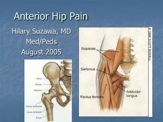
Anterior Knee Pain In Adolescents Johan Myburgh February 2012
Anterior knee pain • Introduction • Case study • Discussion • history • physical examination • investigations • Conditions • Growing skeleton
Introduction • One of the most common musculoskeletal complaints - pediatric population • Differential diagnosis fairly extensive - thorough history and physical examination • Special attention: • anatomic location of the pain • aggravating factors • Assessment of growth and development • Exclude hip and lumbar disorders (all patients)
History • 15 year old male • 2 month history anterior knee pain • Progressively worse • Aggravated by activity • Noticed swelling below knee • Karate – Provincial level • Pain preventing exercise and tournament paticipation
ClinicalExamination • Observation: Swelling at the infrapatellar tendon attachment on the tibial tubercle. • Palpation: Tenderness to same area. • Flexibility: Hamstring tightness • Normal hip and lumbar spine examination
Biomechanicalevaluation • Excessive bilateral subtalar pronation - walking Special investigations: X-ray - fragmentation of the tibial tubercle with overlying soft tissue swelling.
Summary (3 stage) • Clinical. Osgood-Schlatter disease • INTRINSIC FACTORS • biomechanical abnormality • immature skeleton • EXTRINSIC FACTORS • Kicking sport • FITT • Overtraining ( preparing for tournament)
Summary (3 stage) 2. Personal. Karate is his passion - can’t imagine being not able to do it for possibly months. 3. Contextual Couch will not understand the chronic nature of his condition.
Problemlist • Active - Osgood-Schlatter disease • Passive - Excessive bilateral subtalar overpronation
Managementplan • Conservative • Regular icing of the area. • Modifying activities - No pain causing activities like jumping • Physiotherapy to correct biomechanical abnormalities and treat pain. • Progression: • physiotherapy and modified activity routine for 4 weeks • minor relapse of symptoms 2 weeks after resuming sport specific activities, but he started his treatment regime and the pain resolved.
DISCUSSION Anterior Knee Pain
HISTORY • Pain characteristics – location, character, onset, duration, change with activity or rest, aggravating and alleviating factors, and night pain. • Trauma – acute major trauma, repetitive minor trauma. • Mechanical symptoms – locking or extension block, instability • Inflammatory symptoms – morning stiffness, swelling • Bleeding disorders • Previous injury & treatments • Current level of functioning
HISTORY • Overuse knee injuries - report sensation of knee instability • Pseudo-giving way due to a neuromuscular inhibition • Inhibition secondary to pain, muscle weakness and patellar instability.
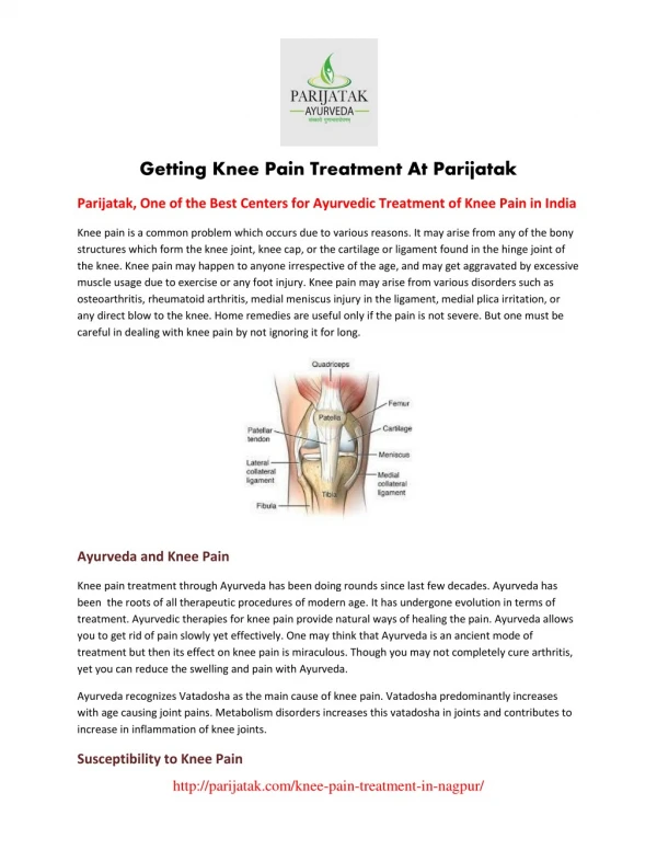
Physical Examination • Complete knee examination (above and below joints) • Examine - contralateral knee and the ipsilateral hip joint. • Biomechanical examination - predisposing factors. • Genetic predisposition includes excessive stiffness, loose-jointedness and poor muscle tone. • Knee joint swelling - suspicion of intra-articular pathology, synovitis
Investigations • Laboratory testing • infection suspected - CBC, ESR, CRP • arthritis is diagnosed - anti-CCP, ANA, RF and HLA-B27 for classification and treatment. • Imaging studies rarely used • Assist in diagnosis • Perthe’s and Slipped femoral capital epiphysis • X-rays and MRI most commonly used.
Extensive differential diagnosis • Patellofemoral pain syndrome • Patellofemoral instability and patellar subluxation • Patellar tendinopathy (Jumper’s knee) • Osteochondroses • Fat pad irritation/impingement • Referred pain from the hip and lumbar spine • Osteochondritis Dissecans • Synovial plica • Quadriceps tendinopathy • Bipartite patella • Stress fracture of the patella • Bursitis • Inflammatory disorders • Pain amplification syndromes • Tumors
Patellofemoral Pain Syndrome • most common cause of pediatric chronic anterior knee pain • etiology • malalignment of the patella relative to the femoral trochlea • result in articular cartilage damage • peripatellar synovitis secondary to mechanical overloading • chemical irritation of local nerve endings
Patellofemoral Pain Syndrome • Risk factors • malalignment of the lower limb • larger Q-angles • VMO weakness • muscle inflexibilities like tight quadriceps, gastrocnemius, hamstrings, lateral retinaculum and IT band. • Classic Hx & Px • Quadriceps grinding test has a 96% sensitivity. • Management • modification of activity, flexibility and strengthening exercises, patellar tracking exercises, icing, NSAIDS, patellar taping and shoe orthotics.
Other patellar pathology • Patellofemoral instability and patellar subluxation • Clinically looks like patellofemoral pain syndrome - but lateral dislocation may be elicited with palpation • Patellar tendinopathy (Jumper’s knee) • common cause of infrapatellar knee pain • associated with osteochondroses and PFP • Rx activity modification and biomechanical rehabilitation • Progressive eccentric strengthening is essential.
OSTEOCHONDROSES • adolescents during growth spurt • present with localized pain with activities , localized tenderness and swelling • X-rays only if infection or bony tumors are suspected. • Self-limiting disorders - managed conservatively • Conservative management includes activity modification, biomechanical rehabilitation, icing, NSAIDS, muscle strengthening and muscle flexibility exercises. • can last ≤ 24 months until skeleton matures. symptoms persist past skeletal maturity surgery indicated to excise the separated tibial tuberosity fragment.
KNEE OSTEOCHONDROSES Patella • Sinding-Larsen-Johansson syndrome (SLJD) Osgood-Schlatter Tibial Tuberosity Tibia • More common • inferior attachment of patellar tendon , epiphysis of the tibial tubercle superior attachment of patellar tendon
OSTEOCHONDROSES Osgood-Schlatter (OSD) Sinding-Larsen-Johansson Syndrome (SLJD)
Osgood-Schlatter Disease • What’s new/controversial ? Journal Pediatrics July 2011 Hyperosmolar Dextrose Injection for Recalcitrant Osgood-Schlatter Disease • injection of the patellar tendon enthesis/tibial apophysis with 12.5% dextrose (monthly x 3) • better 3,6,12 month outcome in pain score (NPPS—Nirschl Pain Phase Scale) than usual care • Release several growth factors and neuropeptides
Conditions • Fat pad irritation/impingement • Infrapatellar fat pad is a richly innervated area • Impingement occurs between the patella and femoral condyle • Caused by direct trauma or a hyperextension injury • Patellar tendinopathy, PFP and synovitis can cause chronic irritation. • Referred pain from the hip and lumbar spine • Perthe’s disease or slipped capital femoral epiphysis may present with knee pain.
Conditions • Osteochondritis Dissecans • Idiopathic bone necrosis • Acute, hemarthrosis and loose body ( locked knee) • Most common lateral aspect of the medial femoral condyle • Synovial plica • Local synovitis caused by microtrauma • synovium trapped between the patella and the femoral condyle. • medial knee pain • a thickened band when pressed against the condyle • Quadriceps tendinopathy • Uncommon
Conditions • Bipartite patella • superolateral patella may show an accessory ossification centre ( pain and swelling) • Stress fracture of the patella • uncommon condition • jumping athletes • intense localized pain and swelling • X-ray chronic stress reaction (bone scan) • Bursitis • Prepatellar bursa most commonly affected • Infrapatellar bursitis mimic tendinopathy • Aspirate bursa if septic arthritis is suspected
Conditions • Inflammatory disorders • Juvenile inflammatory arthritis • morning stiffness and gradual resolution of the pain with activity • monoarthritis • screen for asymptomatic uveitis • confused with OSD (morning symptoms differentiate) • Pain amplification syndromes • Reflex sympathetic dystrophy, reflex neurovascular dystrophy and complex regional pain syndrome • pain out of proportion with the amount of trauma • unwillingness to weight bear and allodynia (pain from a non-painful stimulus) • signs of autonomic dysfunction • special investigations are not helpful.
Conditions • Tumors • rare cause on anterior knee pain • local osteosarcoma, leukemia and metastasis from neuroblastoma
Growing skeleton • Osteochondroses • Referred pain from the hip and lumbar spine • Referred pain form hip and lumber spine
Conclusion • Anterior knee pain - common in the pediatric population • Thorough history and physical examination necessary, often enough to make an accurate diagnosis. • Patellofemoral joint and the extensor mechanism of the knee - most common areas affected • Conditions unique to the growing skeleton like hip diseases (Perthe’s and SCFE) and osteochondroses • Systemic diseases (inflammatory disease and malignancies) should be in differential diagnosis
References • CassasKJ. Childhood and adolescent sports-related overuse injuries. Am Fam Physician. Mar 2006; 73(6): 1014-22. • Patel DR. Musculoskeletal injuries in sports. Prim Care. Jun 2006; 33(2): 545-79. • Mercier LR. Osgood-Schlatter disease. Ferri’s Clinical Advisor: Instant Diagnosis and Treatment. 9th ed. St. Louis, Mo: Mosby; 2009:593 • D Caine, J DiFiori, and N Maffulli. Physeal injuries in children's and youth sports: reasons for concern?, Br J Sports Med. 2006 September; 40(9): 749–760 • Houghton KM. Review for the generalist: evaluation of anterior knee pain. Pediatric Rheumatology 2007, 5:8 • Gastón Andrés Topol, MD,Leandro ArielPodesta, MD, Kenneth Dean Reeves, MD, MarceloFrancisco Raya, PT,Bradley Dean Fullerton, MD,and Hung-wen Yeh, PhD: Journal Pediatrics July 2011 • Brukner and Khan Revised 3rd edition