Download
1 / 10
210 likes | 615 Views
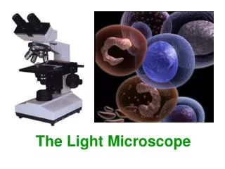
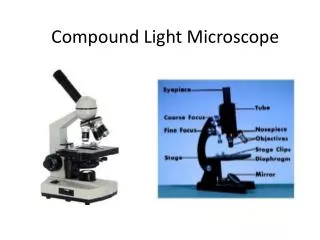
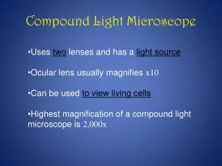
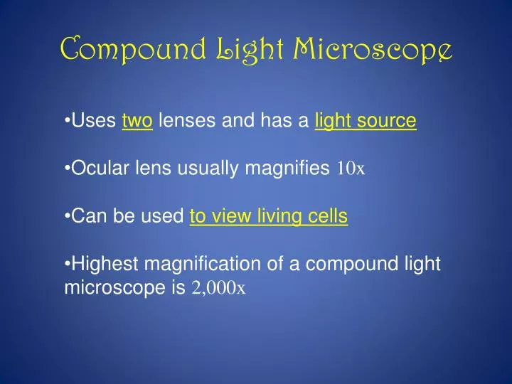
Compound Light Microscope. Uses two lenses and has a light source Ocular lens usually magnifies 10x Can be used to view living cells Highest magnification of a compound light microscope is 2,000x. Electron Microscope. Beam of electrons produce an enlarged image of specimen

E N D
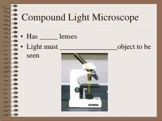
Compound Light Microscope • Uses twolenses and has a light source • Ocular lens usually magnifies10x • Can be used to view living cells • Highest magnification of a compound light microscope is 2,000x
Electron Microscope • Beam of electrons produce an enlarged image of specimen • Highest magnification of an electron microscope is 200,000x • Cannot be used to view living cells
Scanning Electron Microscope • Electron beam is focused on a specimen coated with a very thin layer of metal • Electrons that bounce off form an image on a fluorescent screen • Show 3-D images of cell surfaces • Black and white images only (artificial colors sometimes added)
Transmission Electron Microscope • An electron beam is directed at a very thin slice of a specimen stained with metal ions • Electrons passing through the specimen strike a fluorescent screen, forming an image • The formed image can show the internal structureof a cell in fine detail • Black and white images only (artificial colors sometimes added)
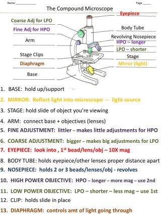
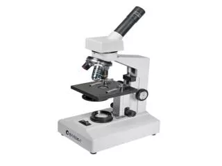
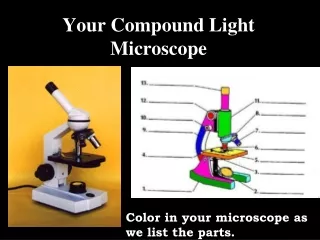
Stage Light Source Ocular Lens Objective Lens10x Objective Lens 4x Objective Lens 40x Course Adjustment Fine Adjustment Stage Clips Base Body Tube Arm Nosepiece Diaphragm/Condenser
Answers Body Tube 12. Coarse Adjustment Nosepiece 13. Fine Adjustment Objective lens 4x 14. Base Objective lens 10x Objective lens 40x Stage Clips Diaphragm / Condenser Light Source Eyepiece Arm Stage
Magnification - increase of an objects apparent size. Resolution - power to show details correctly Contrast - the ability to distinguish different densities of structures Wet Mount - a glass slide holding a specimen suspended in a drop of liquid