Download

1 / 37
590 likes | 1.07k Views
Real time Pcr. Limitations of End-Point PCR. Poor Precision Low sensitivity Low resolution Non - Automated Size-based discrimination only Results are not expressed as numbers Ethidium bromide for staining is not very quantitative Post PCR processing. The Evolution of PCR to Real-Time.

E N D
Limitations of End-Point PCR • Poor Precision • Low sensitivity • Low resolution • Non - Automated • Size-based discrimination only • Results are not expressed as numbers • Ethidium bromide for staining is not very quantitative • Post PCR processing
The Evolution of PCR to Real-Time • Traditional PCRs use Agarose gels for detection of amplification at the final phase or end-point of the PCR reaction • Real time PCR has advanced to allow detection before the end-point of the reaction.
Real-time PCR detection • While the reaction is occurring • monitors the fluorescence emitted during the reaction as an indicator of amplicon production at each PCR cycle • Real-Time chemistries allow for the detection of PCR amplification during the early phases of the reaction
PCR Phases • Exponential: • Exact doubling of product is accumulating at every cycle (assuming 100% reaction efficiency). The reaction is very specific and precise. • Linear (High Variability): • The reaction components are being consumed, the reaction is slowing, and products are starting to degrade. • Plateau (End-Point: Gel detection for traditional methods): • The reaction has stopped, no more products are being made and if left long enough, the PCR products will begin to degrade.
The plateau effect on a fivefold dilution series: • The 5-fold dilution series, seems to plateau at the same place even though the exponential phase clearly shows a difference between the points along the dilution series. • This reinforces the fact that if measurements were taken at the plateau phase, the data would not truly represent the initial amounts of starting target material.
Real-time Principles • Three general methods for the quantitative detection: • Hydrolysis probes (TaqMan, Beacons). • Hybridisation probes (Light Cycler). • DNA-binding agents (SYBR Green).
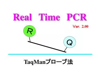
TaqMan Chemistry • Real-time systems using fluorogenic-labeled probes that use the 5´ nuclease activity of Taq DNA polymerase. • Detecting only specific amplification products. • Eliminate post-PCR processing for the analysis of probe degradation
A fluorogenic probe enables the detection of a specific PCR product as it accumulates during PCR. • The TaqMan® Probe is designed with a high-energy dye termed a Reporter at the 5 end, and a low-energy molecule termed a Quencher at the 3 end. • While the probe is intact, the proximity of the quencher dye greatly reduces the fluorescence emitted by the reporter dye by fluorescence resonance energy transfer (FRET) through space. • When reporter and quencher are separated the reporter fluoroscence
How TaqMan Sequence Detection Chemistry Works • If the target sequence is present, the probe anneals downstream from one of the primer sites • During extension the probe is cleaved by the 5´ nuclease activity of Taq DNA polymerase. • The reporter dye is separated from the quencher dye, increasing the reporter dye signal. • The probe is removed from the target strand, allowing primer extension to continue to the end of the template strand.. • Additional reporter dye molecules are cleaved from their respective probes with each cycle.
The fluorescence intensity is increased proportional to the amount of amplicon produced.
Hybridization Probes • Fluorescence Resonance Energy Transfer (FRET) Concept • Donor fluorophore is excited by appropriate wavelength • Donor energy is transferred non-radiatively to the acceptor fluorophore • Excited acceptor emits at longer wavelength • Increase in fluorescence with each cycle • The signal depends only on hybridization
How the Hybridisation probes work? • When the two fluorochromes are in close vicinity (1–5 nucleotides apart), the emitted light of the donor fluorochrome will excite the acceptor fluorochrome (FRET). • This results in the emission of fluorescence, which subsequently can be detected during the annealing phase and first part of the extension phase of the PCR reaction.
SYBR® Green I Dye Chemistry • Small molecules that bind to double-stranded DNA can be divided into two classes: • Intercalators • Minor-groove binders • There are two requirements for a DNA binding dye for real-time detection of PCR: • Increased fluorescence when bound to double-stranded DNA • No inhibition of PCR
How the SYBR Green I Dye Chemistry Works • When SYBR Green I dye is added to a sample, it immediately binds to all double-stranded DNA present in the sample. • During the PCR, the DNA Polymerase amplifies the target sequence, and creates the PCR products. • The SYBR Green I dye binds to each new copy of double-stranded DNA. • As the PCR progresses, more amplicons are created. The result is an increase in fluorescence intensity proportionate to the amount of PCR product produced.
Advantages of SYBR Green I Dye • It can be used to monitor the amplification of any double-stranded DNA sequence. • No probe is required, which reduces assay setup and running costs. • Disadvantage of SYBR Green I Dye • SYBR Green I dye chemistry may generate false positive signals • SYBR Green I dye binds to nonspecific double-stranded DNA sequences. • Needs fully optimized PCR system -no primer dimers or non-specific amplicons. • When not to choose SYBR Green Multiplex reactions.
Terms Used in Quantitation Analysis • Amplification plot: The plot of fluorescence signal versus cycle number
Baseline: The initial cycles of PCR, in which there is little change in fluorescence signal • Passive reference: A dye that provides an internal reference to which the reporter dye signal can be normalized during data analysis. • Rn (normalized reporter): The fluorescence emission intensity of the reporter dye divided by the fluorescence emission intensity of the passive reference dye. • Normalization is necessary to correct for fluctuations caused by changes in concentration or volume.
Rn+: The Rn value of a reaction containing all components, including the template • Rn-:The Rn value of an un-reacted sample. The Rn-value can be obtained from: • The early cycles of a real-time PCR run (those cycles prior to a detectable increase in fluorescence), OR • A reaction that does not contain any template • ΔRn (delta Rn): The magnitude of the signal generated by the given set of PCR conditions. [ΔRn= (Rn+) – (Rn-)]
Threshold: The average standard deviation of Rn for the early PCR cycles, multiplied by an adjustable factor. The threshold should be set in the region associated with an exponential growth of PCR product. • Ct (threshold cycle): The fractional cycle number at which the fluorescence passes the fixed threshold
Quantitation By Real time PCR
Absolute Quantitation • Used to quantitate unknown samples by interpolating their quantity from a standard curve. • Standard: A sample of known concentration used to construct a standard curve. • From a standard curve you can extrapolate the quantity of an unknown sample. • Unknown: A sample containing an unknown quantity of template. This is the sample whose quantity you want to determine
The standard curve resulting from Ct value and standard concentrations should be a linear graph with a high correlation coefficient (> 0.99). • Theoretically a single copy of the target should create a CT value of 40 (if efficiency is 100%), which is the y-intercept in a standard curve experiment • CT value of 40 or more means no amplification and cannot be included in the calculations
Relative Quantitation • A relative quantitation assay is used to analyze changes in gene expression in a given sample relative to another reference sample (such as an untreated control sample). • Relative quantitation might be used to measure gene expression in response to a chemical (drug).
Relative QuantitationStandard curve method • Using a separate standard curve for the target and endogenous control, in separate tubes. • Requires the least amount of optimization and validation.
Relative QuantitationComparative CT method • The need for a standard curve is eliminated. • The target and endogenous control are amplified in the same tube. • Increased throughput: • wells no longer need to be used for the standard curve samples. • the adverse effect of any pipetting and dilution errors made in creating the standard curve samples are eliminated. • A validation experiment must be run to show that the efficiencies of the target and endogenous control amplifications are approximately equal.
Endogenous/Internal Control • Usually an abundantly and constantly expressed housekeeping gene • Best to run a validity test for the selected endogenous control • Combination may be used
Understanding CT • CT (threshold cycle) is the intersection between an amplification curve and a threshold line. • It is a relative measure of the concentration of target in the PCR reaction. • The CT values from PCR reactions run under different conditions or with different reagents cannot be compared directly.
Effect of Efficiency of a PCR Reaction on Ct • The slope of a standard curve is a reflection of the amplification efficiency • The efficiency of the reaction can be calculated by the following equation: Eff=10(-1/slope) –1 • The efficiency of the PCR should be 90-110% (ideal slope = 3.32) • The PCR efficiency is dependent on: • the assay and the master mix performance • sample quality. • length of the amplicon • secondary structure • primer design
Using the PCR Equation Xn = X0(1 + E)n • Xn= PCR product after cycle n • X0= initial copy number • E = amplification efficiency • n = cycle number Xn X0 cycle number
Effect of Amplification Efficiency Xn = X0(1+E)n Case 1: E = 0.9 Case 2: E = 0.8 Xn = 100 (1+0.9)30 Xn = 100 (1+0.8)30 Xn = 2.3 x 10¹⁰ Xn = 4.6 x 10⁹ Result A difference of 0.1 in amplification efficiencies created a five-fold difference in the final ratio of PCR products after 30 cycles
Real-time PCR advantages • Not influenced by non-specific amplification • Amplification can be monitored in real-time • No post-PCR processing of products(high throughput, low contamination risk) • Ultra-rapid cycling (30 minutes to 2 hours) • Most specific, sensitive and reproducible • Not much more expensive than conventional PCR (except equipment cost)