Download
1 / 18
180 likes | 584 Views
Microscopic Anatomy of Selected Male and Female Reproductive Organs. Testes. Each lobule contains one to four seminiferous tubules Tightly coiled structures Function as sperm-forming factories Empty sperm into the rete testis . Figure 16.1. Testes.

E N D
Microscopic Anatomy of Selected Male and Female Reproductive Organs
Testes • Each lobule contains one to four seminiferous tubules • Tightly coiled structures • Function as sperm-forming factories • Empty sperm into the rete testis Figure 16.1
Testes • Each lobule contains one to four seminiferous tubules • Tightly coiled structures • Function as sperm-forming factories • Empty sperm into the rete testis (first part of the duct system) • Sperm travels through the rete testis to the epididymis • Interstitial cells in the seminiferous tubules produce androgens such as testosterone
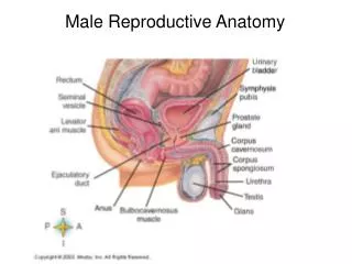
External Genitalia Erections occur when spongy erectile tissue fills with blood during sexual excitement Figure 16.2a
External Genitalia • Internally there are three areas of spongy erectile tissue around the urethra • Erections occur when this erectile tissue fills with blood during sexual excitement
Testes Figure 16.1
The ruptured follicle is transformed into a corpus luteum Ovaries • Each follicle consists of • Oocyte (immature egg) • Follicular cells—surround the oocyte Primary follicle—contains an immature oocyte Graafian (vesicular) follicle—growing follicle with a maturing oocyte Figure 16.7
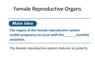
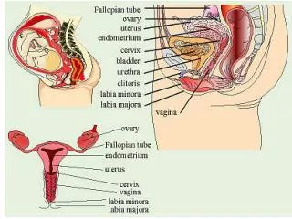
Ovaries • Composed of ovarian follicles (sac-like structures) • Each follicle consists of • Oocyte (immature egg) • Follicular cells—surround the oocyte
Ovarian Follicle Stages • Primary follicle—contains an immature oocyte • Graafian (vesicular) follicle—growing follicle with a maturing oocyte • Ovulation—when the egg is mature, the follicle ruptures; occurs about every 28 days • The ruptured follicle is transformed into a corpus luteum
Hormone Production by the Ovaries • Estrogens • Produced by follicle cells • Cause secondary sex characteristics • Enlargement of accessory organs • Development of breasts • Appearance of axillary and pubic hair • Increase in fat beneath the skin, particularly in hips and breasts • Widening and lightening of the pelvis • Onset of menses (menstrual cycle)
Hormone Production by the Ovaries • Progesterone • Produced by the corpus luteum • Production continues until LH diminishes in the blood • Does not contribute to the appearance of secondary sex characteristics • Other major effects • Helps maintain pregnancy • Prepare the breasts for milk production
Meiotic Events Follicle Developmentin Ovary Before birth 2n Oogonium (stem cell) Follicle cells Mitosis Oocyte Primaryfollicle 2n Primary oocyte Growth Primary oocyte(arrested in prophase I;present at birth) Primaryfollicle 2n (ovary inactive) Childhood Each month frompuberty to menopause Primaryfollicle Primary oocyte (stillarrested in prophase I) 2n Growingfollicle Maturevesicular(Graafian)follicle Meiosis I (completed by oneprimary oocyte each month) Secondary oocyte(arrested inmetaphase II) n First polar body Ovulation Sperm Ovulatedsecondaryoocyte Meiosis II of polar body(may or may not occur) Meiosis II completed(only if spermpenetration occurs) n n n n Polar bodies(all polar bodiesdegenerate) Secondpolar body Ovum Oogenesis Figure 16.10, step 9
Oogenesis and the Ovarian Cycle • Oogonia—female stem cells found in a developing fetus • Oogonia undergo mitosis to produce primary oocytes • Primary oocytes are surrounded by cells that form primary follicles in the ovary • Oogonia no longer exist by the time of birth
Oogenesis and the Ovarian Cycle • Primary oocytes are inactive until puberty • Follicle stimulating hormone (FSH) causes some primary follicles to mature each month • Cyclic monthly changes constitute the ovarian cycle
Oogenesis and the Ovarian Cycle • Meiosis starts inside maturing follicle • Produces a secondary oocyte and the first polar body • Follicle development to the stage of a vesicular follicle takes about 14 days • Ovulation of a secondary oocyte occurs with the release of luteinizing hormone (LH) • Secondary oocyte is released and surrounded by a corona radiata
Ovulation Figure 16.11
Oogenesis and the Ovarian Cycle • Meiosis is completed after ovulation only if sperm penetrates • Ovum is produced • Two additional polar bodies are produced • Once ovum is formed, the 23 chromosomes can be combined with those of the sperm to form the fertilized egg (zygote) • If the secondary oocyte is not penetrated by a sperm, it dies and does not complete meiosis to form an ovum