Download
1 / 17
170 likes | 294 Views
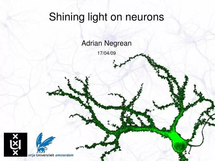
Shining light on neurons. Adrian Negrean 17/04/09. Outline. Neuro-basics Patch-clamping Optical readout of neuronal activity Label-free imaging of live brain tissue. Neuro-basics. distinctive morphology common intracellular components

E N D
Shining light on neurons Adrian Negrean 17/04/09
Outline • Neuro-basics • Patch-clamping • Optical readout of neuronal activity • Label-free imaging of live brain tissue
Neuro-basics • distinctive morphology • common intracellular components • specialized in transducing and conveying information to/from the environment • have mastered the use of ion channels • can modulate their membrane potential
Neuro-basics • equivalent circuit with voltage-dependent ion channels
Patch-clamping credits: Rogier Poorthuis
Optical readout of neuronal activity • Ca2+ imaging is an indirect way of measuring the electrical activity of a neuron • good S/N but slow fluorescence dynamics • focus on membrane potential fluorescent sensors • how does it work ?
Membrane potential sensitive dye at work ANNINE6-plus stained cultured neuron grown on glia, widefield fluorescence imaging The neuron is patch-clamped and the voltage steps are increased gradually Besides the (hopefully) obvious “flickering”, a small and annoying motion artifact is present
Membrane potential sensitive dye at work Neuron grown without glia to suppress background Same staining and imaging as before Membrane potential measurements from different locations
ANNINE6-plus results • the membrane potential is stepped to increasingly depolarized potentials
Nonlinear microscopy tools OPE TPE Two-photon excitation microscopy • excitation in the NIR • low scattering of tissue • deeper imaging • reduced phototoxicity/bleaching • tighter focus
Nonlinear microscopy tools Second and third-harmonic generation microscopy Objective lens incident sample fluorescence SHG THG condenser lens • SHG and THG are forwardly generated • SHG requires non-centrosymmetric media • SHG does not involve excitation of molecular levels, but is enhanced when a two-photon transition can occur • THG is generated at boundaries with a refractive index mismatch
SHG and membrane potential sensitive dyes SHG TPF S-pol. In progress P-pol. Apply intracellularly new dyes and measure their SHG sensitivity to the membrane potential • SF9 cells with extracellular application of FM4-64 dye • SHG at 470 nm, detected with bandpass filter • SHG generated mainly in the outer membrane
Label-free imaging of live brain tissue using THG at 3 x 420 nm (500 x 500 mm) • Cell bodies of neurons • Dead neurons • Blood vessels • “Stuck” red blood cells • Axons and dendrites
Label-free imaging of live brain tissue using THG at 3 x 420 nm (150 x 150 mm) • Nucleus and nucleolus of neurons • Unidentified cellular organelles • Dead neurons • Red blood cells
Label-free imaging of live brain tissue using THG at 3 x 420 nm Sub-cellular structures Red blood cells
Summary • Neurons and electrophysiology • Nonlinear microscopy tools • Membrane potential sensitive dyes • Label-free imaging of live brain tissue with sub-cellular resolution
Acknowledgements and many thanks go to… Prof. Marloes Groot Dr. Stefan Witte Dr.ir. Erwin Peterman Prof. Johannes de Boer Dr. Mattijs de Groot Assist. Prof. Ruud Toonen Prof. Huibert Mansvelder Hans Lodder • as well as the other people from • the “Neuro-Laser” think-tank • my supervisors • and many thanks to my colleagues from the electrophysiology dept.