Download
1 / 28
280 likes | 528 Views
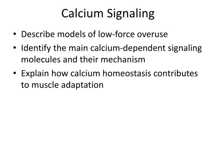
Calcium Signaling. Describe models of low-force overuse Identify the main calcium-dependent signaling molecules and their mechanism Explain how calcium homeostasis contributes to muscle adaptation. Low force overuse. Models Chronic stimulation Endurance training Physiological stresses

E N D
Calcium Signaling • Describe models of low-force overuse • Identify the main calcium-dependent signaling molecules and their mechanism • Explain how calcium homeostasis contributes to muscle adaptation
Low force overuse • Models • Chronic stimulation • Endurance training • Physiological stresses • Electrophysiological • Oxygen delivery/handling • ATP metabolism • Adaptation • SR swelling • Mitochondrial hypertrophy • “Slow” phenotype expression • Atrophy
Acute changes during contraction • Phosphate redistribution • pCrATP • ATP2 Pi + AMP • pH decline 2 Hz 10 Hz Time (min) Kushmerick & al., 1985
Changes in blood composition 5 min exercise 10 min recovery • Lactate appears ~3 min • pH falls in parallel • Norepinepherine Gaitanos &al 1993
Glucose and FFA liberation • 70% VO2 max, 2h • Muscle glycogen falls • Energetic molecules released from non-muscle stores Krssak & al 2000
Calcium redistribution • Mitochondrial • Rise ~2x during exercise • Remains elevated > 1 hour • Cytoplasmic • Spikes to 1 uM (diminishing) • Baseline to 300 nM • Metabolite imbalance exceedsexercise period Rest 60 min Ex 30 min Rest 60 min Rest Madsen & al., 1996
Stimulation-dependent signaling • Calcium • Troponin/tropomyosin: contraction • Calcineurin: gene transcription • Calpain: structural remodeling • CaMK: transcription, channel activity • Energy/ATP • AMP kinase: glucose transport, protein balance • PPAR: mitochondrial hypertrophy • ROS: complicated
Chronic electrical stimulation • Stanley Salmons & Gerta Vrbova, 1969 • Spinal-isolated & tenotomized soleus • ie: no voluntary or reflex activation • Normally highly active muscle • Stimulate 1-40 Hz, 67% duty cycle 8 hr/day • Implanted stimulator tibialis anterior • 24/7, 10 Hz • Normally low activity muscle
Stim frequency contraction time • Soleus (slow muscle) • Tenotomyatrophy • Tenotomyfaster • Tenotomy+low frequency preserve speed • Tenotomy+high frequency faster • Stimulation frequency influences • Calcium kinetics • Troponin kinetics • Myosin kinetics Normal 10 Hz 40 Hz
Stim frequency contraction time • TA (fast muscle) • No tenotomy no atrophy • Stim effects • Slower • Reduce Twitch-tetanus ratio • Reduce sag 10 Hz Twitch forces Tetanic forces
Mechanical performance changes • P0 declines (atrophy) • Vmax declines (slower) • Endurance increases 2 weeks CLFS Control muscle Jarvis, 1993
Structural adaptation Normal • Reduced T-tubules • Wider Z-lines • More mitochondria • More capillaries Stimulated Z-line width Stimulation Recovery Eisenberg, 1985
Endurance training • Typically 6 weeks, 5/week 30-120 min @ 60-80% VO2max • Performance & oxygen adapts • Contractile proteins less so Pre-train 6 wks 6 mos Heart Rate Lactate Power (watt) Hoppeler & al 1985
Endurance adaptation paradigm • Elevated calcium and AMP activate mitochondrial genes • AMPK, PGC-1, pPAR, MEF2 • Elevated calcium activates muscle genes Baar, 2006
Ca mediated protein modification • CaMK (I – IV) • Calmodulin mediated • Serine/threonine kinases • CaMK-III = eEF2 kinase • Post-synaptic density • Protein kinase C • Calcineurin • Calmodulin mediated • Serine/threonine phosphatase • Calpain (I-III) • Cysteine protease • Cytoskeletal remodeling
Calcium controls everything http://www.genome.jp/kegg-bin/show_pathway?hsa04020
Calcineurin (Cn) • Calcium & calmodulin dependent • Serine/threonine phosphatase • High calcium sensitivity: 200 nM • Transcriptional targets • NFAT • MEF2 • Functional targets • DHPR • BAD CnB CnA CaM Li & al., 2011
MEF2 • MEF2 A/C/D “MADS-box” transcription factor • Compliment myogenic regulatory factors • Cn and p38-dependent • Blocked by class 2 HDACs • MHC, MLC, Tm, Tn • NADH dehydrogenase (complex 1), GLUT4 Activation Domain: HDAC/MRF interactions MEF2 protein map (NLM)
NFAT • Stimulation-dependent nuclear translocation • 30 minutes, 10 Hz; recovery Liu & al 2001
NFAT • NFAT 1/2/3/4 transcription factor • MEF2, AP-1 cooperation • Cn, GSK3, PKA dependent • Sensitive to mitochondrial calcium handling • Myoglobin, TnI(slow), MHC(slow) NFAT protein map (NLM)
SURE and FIRE • Slow Upstream Regulatory Element (SURE) • Identified in TnI-slow • 110 bp, contains both MEF2, E-box, GT-box • Fast Intronic Regulatory Element (FIRE) • Identified in TnI-fast • 150 bp in Intron 1, MEF2, E-Box, GT-box • NFAT-binding • Upstream: promoter • Intron: repressor
HDAC • Histone deacetylase : gene inactivation • HDAC 2-5; Sirt • MEF2 compliment • CaMK/PDK1 phosphorylation • Nuclear export • 14-3-3 binding • ie: blocks MEF2-mediated transcription when not phosphorylated
Activity dependent transcription Frequent activity Infrequent activity Low Resting Calcium Transient Calcium Spike High Resting Calcium Cn Inactive CaMK Active Cn Active MEF2 HDAC2 Myosin Myoglobin NFAT NADH-D Actin
CaMKII autophosphorylation • CaM Kinase II (CaMKII) • CaM dependent kinase • CaM kd = 2 nM, koff 0.3/s • High affinity, fast kinetics • Phospho-CaMKII • CaM independent kinase • CaM kd = 0.1 pM, koff 10-6/s • Very high affinity, slow kinetics • CaMKII autophosphorylation locks itself in an active conformation
Rate decoding • Autophosphorylation is like integration • Dephosphorylation is like a high pass filter • eg: Deliver regular calcium pulses • Measure Ca independent activity • Elevated > 1 hr after exercise in muscle
CaMK effectors • MEF2 • CREB • CBP/p300 Histone Acetyltransferase partner • Creatine Kinase, SIK (HDAC) • PGC-1a • Carnitine palmitoyltransferase • Mitochondrial transcription factor A (Tfam)
VEGF • Vascular Endothelial Growth Factor • Angiogenesis
Summary • Sustained contractile activity disrupts calcium and ATP homeostasis • Calcium-dependent kinases (CaMK) and phosphatasis (Cn) alter transcription (MEF2, NFAT, PGC1) • Altered gene expression results in mitochondrial biogenesis and calcium buffering • Subsequent activity causes less disruption