Download
1 / 9
1.54k likes | 11.56k Views
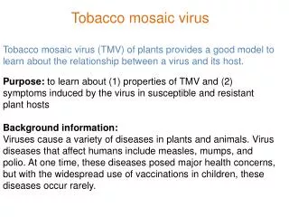
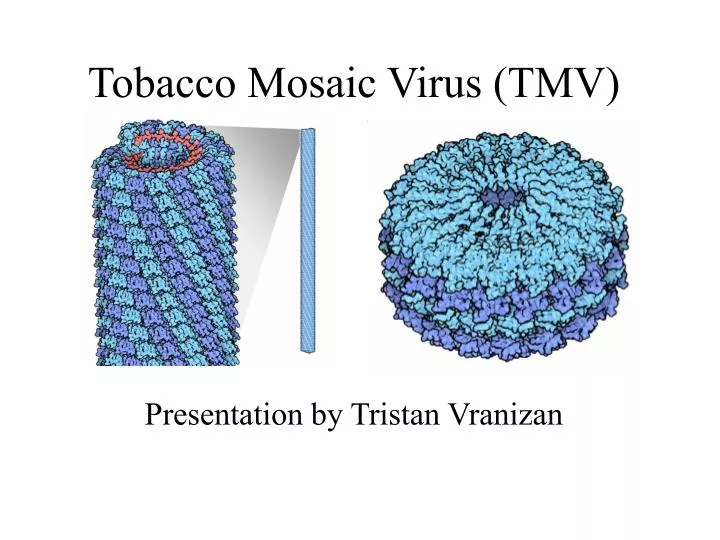
Tobacco Mosaic Virus (TMV). Presentation by Tristan Vranizan. History. TMV: the first virus to be discovered Late 19th Century: Researchers found out that a tiny infectious agent was the cause of a disease that was killing tobacco plants.

E N D
Tobacco Mosaic Virus (TMV) Presentation by Tristan Vranizan
History • TMV: the first virus to be discovered • Late 19th Century: Researchers found out that a tiny infectious agent was the cause of a disease that was killing tobacco plants. • Wendell Stanley coaxed the virus to form crystals and found out that it was mainly composed of proteins. • RNA (Ribonucleic acid) was also discovered in the virus Wendell Stanley >> Won Nobel prize for his study.
Description of Molecule • TMV has one strand of RNA, shown in red. • RNA is wrapped with stacks of protein, shown in blue. • Protein Coat: 2,130 copies of a small protein stack on top of each other, spiraling downward. • RNA strand is encoded in four proteins, transporting the RNA from cell to cell and allowing the virus to spread.
Infection • TMV is so stable it can survive in cigars for years made from infected tobacco leaves. • The coat of proteins, around the RNA strand, protect it from any enzymes trying to kill it. • But, in order for TMV to spread it has to release it’s coat of proteins for the RNA to enter a cell • The proteins, situated outside the cell, repel against each other. This allows the cell to release RNA back into the coat of proteins to spread again. Healthy tobacco leaf > < TMV infected leaf
Assembly • Mixing RNA with capsid proteins creates a functional virus. • Two steps create this process • Step 1: Proteins form into a two-layer disk, RNA then fills the hole at the center causing the proteins to form into a ring, eventually covering the RNA. • Step 2: RNA is ensured to being packaged into viruses with its’ need for an initiation sequence.
Structure • 17 subunits surround the RNA in red. • RNA is located in the grooves of the protein, wedged between them. • 3 nucleotides bind each protein subunit.
Structure (continued) • The bright red dots are the acidic amino acids. • They help to break up the virus inside infected cells.
Picture References • http://erec.ifas.ufl.edu/tomato-scouting-guide/images/diseases/tobacco-mosaic135.JPG • http://erec.ifas.ufl.edu/tomato-scouting-guide/diseases/tobacco-mosaic-virus.shtml • http://matyasciprian.hu/kepek/paprika_betegsegek/paprika_tmv_3.JPG