Download
1 / 12
130 likes | 507 Views
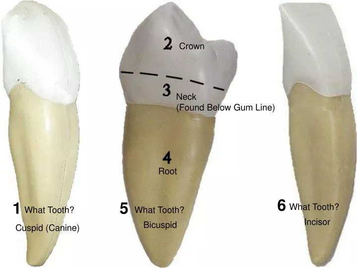
Crown. Neck (Found Below Gum Line). Root. 6. 1. 5. What Tooth?. What Tooth?. What Tooth?. Incisor. Bicuspid. Cuspid (Canine). Enamel. 1. Dentin. 2. Pulp. 5. Cementum and Periodontal Ligament attach on this surface. 3. 6. What Tooth?. Pulp in Root Canal. Lower Molar

E N D
Crown Neck (Found Below Gum Line) Root 6 1 5 What Tooth? What Tooth? What Tooth? Incisor Bicuspid Cuspid (Canine)
Enamel 1 Dentin 2 Pulp 5 Cementum and Periodontal Ligament attach on this surface 3 6 What Tooth? Pulp in Root Canal Lower Molar (Upper Molar has three Roots) 7 Root 4 Root Canal
Internal Nares Superior Nasal Conchae Middle Nasal Conchae 1 Opening ofEustachian Tube 2 Inferior Nasal Conchae Nasopharynx 13 3 15 14 Pharyngeal Tonsils Hard Palate 4 16 5 Soft Palate 6 Palatine Tonsils 17 Uvula 18 Oropharynx 7 19 Sublingual Gland 8 Fauces 9 20 Submandibular Gland Laryngopharynx Epiglottis 10 11 Ventricular Folds Vocal Folds 21 12 Cricoid Cartilage Thyroid Cartilage
Falciform Ligament Left Liver Lobe Right Liver Lobe Body of Stomach Transverse Colon Jejunum Greater Omentum 7 Mesentery Proper Ileum
No 1,4,7 Fundus Lower Esophageal (Cardiac) Sphincter Beginning of Duodenum Lesser Curvature Cardiac Region Rugae Pyloric Canal Pyloric Sphincter Body of Stomach Pyloric Antrum Greater Curvature 13 Head of Pancreas
ABC first, then #’s Ampulla of Vater opening Muscularis Externa Accessory Duct Pancreatic Duct 1 Spleen Pancreas 6 7 2 3 5 4 Plica Circularis Beginning of Jejunum Duodenum Superior Mesenteric Artery
ABC first, then #’s Hepatic Portal Vein Common Bile Duct Hepatic Artery Spleen Celiac Artery 2 1 3 4 Superior Mesenteric Artery Pancreas Duodenum
2 Gall Bladder Right Hepatic Duct Round Ligament (free edge of the falciform ligament) 3 1 Right Lobe of Liver 6 8 Left Hepatic Duct Cystic Duct of Gall Bladder 5 9 Hepatic Artery Common Hepatic Duct 7 Hepatic Portal Vein 10 4 Left Lobe of Liver Inferior Vena Cava 11
2 Pancreas Duodenum 1 3 Pancreatic Duct Ascending Colon 4 Taenia Coli Transverse Colon 5 5 7 6 Haustra 8 Descending Colon 9 Rectum Anal Canal 10
Pancreas Duodenum 2 Pancreatic Duct 1 Mucosa of Duodenum 3 Beginning of Jejunum 5 4 6 13 Ascending Colon Descending Colon 6 10 7 13 Ileocecal Valve 8 End of Ileum 9 Cecum 11 Appendix 14 Sigmoid Colon 11 Taeniae Coli 12 Rectum Anal Canal 15
Villi Villi Capillaries Lacteal Crypts of Lieberkuhn (Intestinal Glands) Lamina Propria Muscularis Mucosae Lymphatic Nodule Brunner’s (Duodenal Mucous) Gland Submucosal Plexus 6 Myenteric Plexus Circular Layer of Muscularis Externa Logitudinal Layer of Muscularis Externa Simple Squamous Epithelium of Serosa 8
Lacteal Simple Columnar Epithelium of Mucosa Goblet Cells