Download
1 / 29
520 likes | 1.65k Views
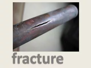
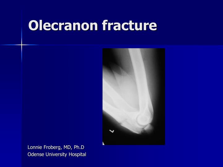
Olecranon fracture. Lonnie Froberg , MD, Ph.D Odense University Hospital. 20% of forearm fracture 12 per 100.000 persons per year Low-energy fall Increased risk >50 years 90% AO 21.B1.1. Duckworth et al. Injury 2012;43:343-346. Why operate? Methods of fixation K-wire, cerklage

E N D
Olecranon fracture Lonnie Froberg, MD, Ph.D Odense University Hospital
20% of forearm fracture • 12 per 100.000 persons per year • Low-energy fall • Increased risk >50 years • 90% AO 21.B1.1 • Duckworth et al. Injury 2012;43:343-346
Why operate? • Methods of fixation • K-wire, cerklage • Plating • Outcome • Summary
Why operate? • Restore articular surface • Achieve absolute stability • Commence early active movement • Preservation of range of motion and power • Avoidance of complications
Methods of fixation? • Cadaveric elbow joint • Standard osteotomies • Five different fixation techniques • Loads applied comparable to clinical situations • Displacements measured Fyfe et al. Jour Bone Joint Surg (Br).1985. 67B;3:367-372
Methods of fixation? Fyfe et al. Jour Bone Joint Surg (Br). 1985. 67B;3:367-372
Methods of fixation? Fyfe et al. Jour Bone Joint Surg (Br). 1985. 67B;3:367-372
K-wire with or without eyelets? • No significant difference in postoperative pain or in rate of hard ware removal Kim et al. J Hand Surg Am. 2013.Jul 9
How to place the K-wires? • Proximal ulnar canal? • Anterior cortex? • Distal ulnar canal? Huang et al. J Trauma. 2010.68;1:173-176
How to place the K-wires? • Inserted as close as possible to the articular surface • Back 1 cm from final position, cut obliquely, bent • Incisions with lines in triceps • K-wires are impacted into ulna Newman et al. 2009. Injury; 40(6): 575-581
How to place the K-wires? • K-wire penetration more than 10 mm beyond the anterior cortex increases risk for penetration of median nerve and ulnar artery Prayson et al. Shoulder Elbow Surg. 2008.17;1:121-125
Which kind of tension band? Lalliss et al. Jour Bone Joint Surg (Br).2010.92B;2:315-319
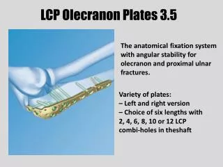
Plating • When to plate? • Tension band is not appropriate • Oblique fractures distal to the midpoint of the troclear notch • Co-existing coronoid fracture • Associated with Monteggia fracture dislocation Newman et al. 2009. Injury; 40(6): 575-581
Which kind of plate? • Cadaveric study • Comminute fracture • No difference in failure rate (>2 mm gap of fracture) Buijze et al. Arch Orthop Trauma Surg.2010;130:459-464
Which kind of plate? • Advantage of locking compression plate to conventionel plate: • Angular and axial stability • Preserves periosteal blood supply • No toggling of unlocked screws (improves fixation in osteoporotic fractures and comminution)
Which kind of plate? • Stainlesssteel or titanium? • More screw in proximal fragment betterthanfewerscrews? • Largerscrewsbetterthan small screws?
Which kind of plate? • Accumedstainlessstell • Synthesstainlessstell • Synthes titanium • US Implants • Zimmer • Edwards et al. J Orthop Trauma 2011;25(5):306-311
Which kind of plate? • No statistical difference between maximum load and cycles survived • Edwards et al. J Orthop Trauma 2011;25(5):306-311
Outcome – Cochrane review Veillette et al. OrthopClin N Am. 2008;39:229-236
Summary – Tension band fixation • Fracture: Transverse or oblique • K-wire: Anterior cortex or distal ulnar canal • K-wire penetration: <10 mm beyond the anterior cortex • Tension band: 1.0 mm stainless steel wire, 2 knots
Summary - Plating • Fractures: Distal to the midpoint of the troclear notch, co-existing coronoid fracture, Monteggia • Locking compression plate theoretically superior to conventionel plate