Download
1 / 1
20 likes | 111 Views
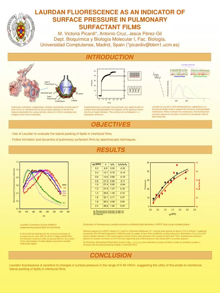
50. Laurdan molecule. 40. Ordered Phase. 30. Pressure. 20. Type II Pneumocyte. Disordered Phase. 10. 0. Surfactant. Interstitial layer. Air space. Type I Pneumocyte. 0. 5. 10. 15. 20. 25. 30. Capillar. Time. Endothelium. Fluorescence Intensity. . 490. 440.

E N D
50 Laurdan molecule 40 Ordered Phase 30 Pressure 20 Type II Pneumocyte Disordered Phase 10 0 Surfactant Interstitial layer Air space Type I Pneumocyte 0 5 10 15 20 25 30 Capillar Time Endothelium Fluorescence Intensity 490 440 Laurdan´s emission is blue-shifted in progressively packed lipid environments. In films with low lipid density the maximum emission of Laurdan occurs near 490 nm while in highly packed films the emission maximum shifts to around 440 nm, as a result of the reorientation of water dipoles around the excited-state probe dipole. Evaluation of fluorescence Laurdan emission at different lipid densities in DPPC films using multiwell plates. Different aliquots of a DPPC solution 0,1 mg/ml in Chloroform/Methanol (2:1, vol/vol) were spread on top of a Tris 5 mM pH 7 subphase containing 150 mM NaCl deposited in different wells of a plate, to form films at different surface pressures. Afterwards, 2-5 mL of a 0,07 mg/ml Laurdan solution in the same organic solvent mixture was spread on the surface of each well. Then, fluorescence emission spectra of Laurdan (excitation at 370 nm) was registered and surface pressure was measured in a surface balance. An Excitation Generalized Polarization function (Gpex: IB-IR/IB+IR) was selected to analyze all data in order to correlate Laurdan´s emission with the lateral packing of lipids in interfacial films. LAURDAN FLUORESCENCE AS AN INDICATOR OF SURFACE PRESSURE IN PULMONARY SURFACTANT FILMSM. Victoria Picardi*, Antonio Cruz, Jesús Pérez-GilDept. Bioquímica y Biología Molecular I, Fac. Biología, Universidad Complutense, Madrid, Spain (*picardiv@bbm1.ucm.es) INTRODUCTION Laurdan (6-Lauroyl-2-(N,N-dimethylamine) naphtalene) is a fluorescent probe used to analyse the structure of phospholipid membranes. When inserted into membranes, the fluorescence emission spectra of Laurdan is sensitive to the phase state of lipid molecules. Pulmonary surfactant, a lipid-protein complex secreted by alveolar type II cells, forms an interfacial film at the air-liquid interface of alveoli that reduces drastically the surface tension (close to 0 mN/m) avoiding lung collapse at the end of expiration. A good pulmonary surfactant must promote very rapid transfer of surface active phospholipids from bilayers at the aqueous phase into the air-liquid interface. This activity can be followed in -t adsorption isotherms. OBJECTIVES - Use of Laurdan to evaluate the lateral packing of lipids in interfacial films. - Follow formation and dynamics of pulmonary surfactant films by spectroscopic techniques. RESULTS mN/m 8.9 21.3 12.6 27.4 32.1 38.0 IB: Fluorescence Intensity at 445 nm IR: Fluorescence Intensity at 490 nm : Pressure CONCLUSION Laurdan fluorescence is sensitive to changes in surface pressure in the range of 0-40 mN/m, suggesting the utility of this probe to monitorize lateral packing of lipids in interfacial films.