Download
1 / 38
400 likes | 1.33k Views
Mechanism of action of anti- trypanosomal drugs. Suramin:. Suramin inhibits trypanosome glycolytic enzymes more effectively that it does those of the host . Melarsoprol: Similar to suramin, melarsoprol also inhibits enzymes of glycolytic pathway . Pentamidine:

E N D
Mechanism of action of anti-trypanosomal drugs Suramin: Suramin inhibits trypanosome glycolytic enzymes more effectively that it does those of the host Melarsoprol: Similar to suramin, melarsoprol also inhibits enzymes of glycolytic pathway Pentamidine: Pentamidine binds mitochondrial DNA of trypanosomes (kDNA) and disrupts its function 1
Armadillo, opossum, raccoon, dog cat, and other animals T. cruzi:life cycle Reservoir 4
Triatoma sordida, T. infestans, Panstrongylus megistus, Rhodnius prolixus, etc. kissing bugs cone nose bugs 6
Stage Stage Symptoms Symptoms Organ involved Organ involved Primary lesion Nonpurulent (without pus) edematous (swollen with an excessive accumulation of fluid) plaque. Skin Malaise, restlessness, lymphadenitis (inflammation of lymph node), hepatomegaly, splenomegaly, acute myocarditis, generalized edema Acute stage Lymph node and heart Chronic stage Hollow organs Mega esophagus, mega colon, cardiomegaly, cardiac arrhythmia Chagas’ disease: symptoms 8
Chagas’ disease: Symptoms (chagoma) 9
Chagas’ disease: mega colon 10
Chagas’ disease: mega colon 11
Chagas’ disease: megacardium 12
Chagas’ disease: T. Cruzi diagnosis Trypanosoma cruzi parasite in a thin blood smear. (CDC Photo) 13
Direct Inflammatory response • damage to infected cells • Destruction of autonomic nerve ganglions • Products of humoral and CMI 14
Antibody kills, but not eradicate T. cruzi • CMI of some protective value • Activation macrophages kill the organism • Immunosuppression 15
Chagas' disease:diagnosis • History of travel and sometimes cardiac problems • Romana’s sign or chagoma • Organisms in the chagoma exudate, lymph node aspirate or blood 16
Use of Polymerase Chain Reaction to Diagnose the Fifth Reported US Case of Autochthonous Transmission of Trypanosoma cruzi, in Tennessee, 1998 In July 1998, the mother of an 18-mo.-old boy in rural Tenn. found a triatomine bug in his crib, which she saved because it resembled a bug shown on a TV program about insects that prey on mammals. The gut contents of the bugwere found, by light microscopy and PCR, to be infected with T. cruzi. Blood specimens obtained from the child in July and August were negative by microscopy and hemoculture but positive by PCR and DNA hybridization, suggesting a low level parasitemia. Specimens obtained after benznidazole treatment were negative. He did not develop anti–T. cruzi antibody; 19 relatives and neighbors also were seronegative. Two of 3 raccoons trapped in the vicinity had positive hemocultures for T. cruzi. (J. Infect. Dis. 2000;181:395–9) 18
Trypanosoma cruzi in Transplant recipients Case 1. In December 2005, a man aged 64 years received a heart transplant. In February 2006, he was readmitted to the hospital with anorexia (eating disorder), fever, and diarrhea of 2 weeks' duration. T. cruzi was seen in both blood and biopsy from the heart. The patient had no identifiable risk factors for T. cruzi infection (e.g., travel to a country endemic for Chagas disease). He was seronegative for T. cruzi antibodies but positive for T. cruzi DNA by polymerase chain reaction (PCR). After initiation of nifurtimox therapy, his parasitemia rapidly cleared. However, in April 2006, the patient died from complications attributed to acute rejection of the transplanted organ. The source of infection tracked the organ donor who tested seropositive for T. cruzi. antibodies by RIPA (radioimmunoprecipitation assay) and tested borderline-positive by IFA (immunofluorescent antibody). He had been born in the US but had traveled to a T. cruzi-endemic area of Mexico. 19
Trypanosoma cruzi in Transplant recipients Case 2. In January 2006, a man aged 73 years received a heart transplant. The patient was readmitted to the hospital in February 2006 with fever, fatigue, and an abdominal rash. A thin blood smear revealed T. cruzi trypomastigotes, and blood cultures were positive for T. cruzi. The patient had no identifiable risk factors for T. cruzi infection. He was seronegative but PCR-positive for T. cruzi, indicating recent infection. The patient's rash and parasitemia resolved after 10 days of nifurtimox treatment. Serial endomyocardial biopsies did not reveal trypanosomes, and he remained seronegative by IFA for T. cruzi. The patient died in June 2006 from a cardiac failure. The source of infection was the organ donor, who had been born in El Salvador and was residing in Los Angeles at the time of his death, tested positive for T. cruzi antibodies. 20
Nifurtimox effective in acute stage • Benznidazole may be of value in chronic stage • Avoid and control the insect population • No vaccine 21
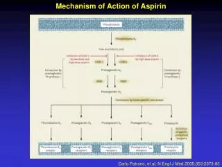
Nifurtimox- generates cytotoxic superoxide anion (02-) which is toxic to the parasite Benznidazole- protein- and RNA-synthesis inhibitor 22
Leishmaniasis Organism Disease Epidemiology L. donovani Asia: 10x106 visceral leishmaniasis L. tropica cutaneous leishmaniasis Mediterranean: 5x10⁶ L. Braziliensis and other mucocutaneous leishmaniasis South/Central America: 10x106 24
Leishmaniamorphology • Amastigote (leishmania) seen in the mammalian host • Promastigote (leptomonad) seen in sand fly 26
Leishmanialife cycle Human cycle Animal reservoir 27
Sand fly vector Leishmania life cycle 28
Type Organ Involved Symptoms Visceral Liver, spleen, bone marrow, lymph nodes, skin No bite reaction; lymphadenopathy, splenomegaly and hepatomegaly; parasitemia, chills and fever; darkening of skin Cutaneous Skin Centrifugally growing papular lesion with central crusting; heals spontaneously, permanent scar Muco-cutaneous Skin and mucoid tissue Initially same as cutaneous lesion but it does not heal: necrosis of mucoid tissue; metastasis to distant mucoid tissues; very disfiguring 29
1-4 months: fever chills, diarrhea, dysentery • Progressive hepato-splenomegaly • Skin hyperpigmentation • Death, if untreated 30
Patient A. Fever (104º F) was first noted by patient A in late December 2003 and rigors, flushing, sweats, and mild orthostasis in early January 2004 and lost 13 pounds of body weight. Visceral Leishmaniasis was suspected but no leishmanial parasites were noted on lightmicroscopic examinations or cultures of bone-marrow and liver-biopsy specimens, and no leishmanial DNA was detected by genus-specific polymerase chain reaction (PCR). The findings in the splenic enlargement suggested, and surgical splenectomy was considered briefly. In February 2004, because of continuing concern that the patient had VL, the liver-biopsy specimen was reexamined by light microscopy; one definite and multiple probable leishmanial parasites were noted. 31
Visceral Leishmania: recent cases Patient B. Abrupt onset of fever 104º F, myalgia, and abdominal pain were noted in mid-December 2003. Symptoms worsened and there was anorexia, with a loss of 25 pounds of body weight) during the next 6 weeks. No leishmanial parasites were found on light microscopic examinations of bone-marrow and buffy-coat specimens but were prevalent in a liver-biopsy specimen. During February 3–17, 2004, the patient was treated with a lipid formulation of amphotericin B, resulting in a transient improvement of symptoms. He was rehospitalized on March 5, with a temperature of 102º F. Results of PCR analysis indicated Leishmania donovani. On March 19, a 28-day course of antileishmanial therapy was begun with Pentostam® (dose: 20 mg/kg/d, intravenously 32
Leishmania: Diagnosis • History • Lesions or symptoms • Organisms in the lesion • Montenegro test (type IV hypersensitivity) 35
LeishmaniasisPathogenesis and immunology • Damage due to CMI • Leishmanialproteoglycan • Leukopenia with monocytosis and lymphocytosis • Immunosuppression • Interferon and TNF are protective 37
LeishmaniasisPreventions and treatment • No vaccine • Control of sand fly and infected animals • avoidance of sand fly • Pentosam (antimony gluconate) 38