Download
1 / 84
880 likes | 1.29k Views
Fundamentals of the Nervous System and Nervous Tissue. Chapter 11. Introduction. The nervous system is the master controlling and communicating system of the body It is responsible for all behavior

E N D
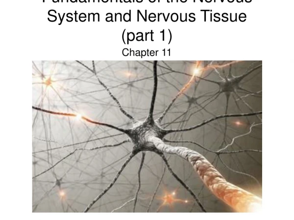
Fundamentals of the Nervous System and Nervous Tissue Chapter 11
Introduction • The nervous system is the master controlling and communicating system of the body • It is responsible for all behavior • Along with the endocrine system it is responsible for regulating and maintaining body homeostasis • Cells of the nervous system communicate by means of electrical signals
Nervous System Functions • The nervous system has three overlapping functions • Gathering of sensory input • Integration or interpretation of sensory input • Causation of a response or motor output
Introduction • Sensory input • The nervous system has millions of sensory receptors to monitor both internal and external change • Integration • It processes and interprets the sensory input and makes decisions about what should be done at each moment • Motor output • Causes a response by activating effector organs (muscles and glands)
Organization • There is only one nervous system; however, for convenience the nervous system is divided into two parts • The central nervous system • Brain and spinal cord • Integrative and control centers • The peripheral nervous system • Spinal and cranial nerves • Communication lines between the CNS and the rest of the body
Organization • The peripheral nervous system has two fundamental subdivisions • Sensory (afferent) division • Somatic and visceral sensory nerve fibers • Consists of nerve fibers carrying impulses to the central nervous system • Motor (efferent) division • Motor nerve fibers • Conducts impulses from the CNS to effectors • (glands and muscles)
Organization • The motor division of the peripheral nervous system has two main subdivisions • The somatic nervous system • Voluntary (somatic motor) • Conducts impulses from the CNS to skeletal muscle • The autonomic nervous system (ANS) • Involuntary • Conducts impulses from the CNS to cardiac muscles, smooth muscles, and glands
Organization • The autonomic nervous system has two principle subdivisions • Sympathetic division • Mobilizes body systems during emergency situations • Parasympathetic division • Conserves energy • Promotes non-emergency functions • The two subdivisions bring about opposite effects on the same visceral organs • What one subdivision stimulates, the other inhibits
Peripheral Nervous System • Visceral organs are served by motor fibers of the autonomic nervous system and by visceral sensory fibers • The somata (limbs and body wall) are served by motor fibers of the somatic nervous system and by sensory somatic sensory fibers • Arrows indicate the direction of impulses
Histology of the Nervous Tissue • Nervous tissue is highly cellular • Less that 20% of the CNS is extracellular space • Cells are densely packed and tightly intertwined • Nervous tissue is made up of two cell types • Neurons • Excitable cells that transmit electrical signals • Support cells • Smaller cells that surround and wrap the delicate neurons • These same cells are found within CNS and PNS
Supporting Cells • All neurons associate closely with nonnervous support cells of which there are 6 types • Support cells of the CNS • Astrocytes • Microglial • Ependymal • Oligodendrocyte • Support cells of the PNS • Schwann cells • Satellite cells
Supporting Cells in the CNS • The supporting cells of the CNS are collectively called neuroglia or simply, glial cells • Like neurons, glial cells have branching processes and a central cell body • Neuroglia can be distinguished by their much smaller size and by their darker staining nuclei • They outnumber neurons in the CNS by a ration of 10 to 1 • Make up half of the mass of the brain
Astrocytes • Star shaped • Most abundant type of glial cell • Radiating projections cling to neurons and capillaries, bracing the neurons to their blood supply • Astrocytes play a role in exchanges between capillaries and neurons
Astrocytes • Cells function as antigen presenting cells of the immune response • Control chemical environment around neurons, recapturing potassium ions and released neuro- transmitters • Astrocytes signal each other via intracellular calcium pulses
Microglial • Small ovid cells with relatively long “thorny” processes • Their branches touch nearby neurons to monitor health of the neuron • Microglial migrate toward injured neurons
Microglial • Small ovid cells with relatively long “thorny” processes • Their branches touch nearby neurons to monitor health of the neuron • Microglial migrate toward injured neurons
Microglial • When invading micro- organisms are present or damaged neurons have died, the micro- glial transforms into a special type of macro- phage that protects the CNS by phagocytizing the microorganisms or neuronal debris • Important because cells of the immune system can enter CNS
Ependymal • Range in shape from squamous to columnar and many are cilated • Line the central cavities of the brain and spinal cord • Form a fairly permeable barrier between cerebrospinal fluid of those cavities and the cells of the CNS • Beating cilia circulates cerebrospinal fluid
Oligodendro- cytes • Fewer branches than astrocytes • Cells wrap their cytoplasmic extensions tightly around the thicker neurons in the CNS • Produce insulating coverings called myelin sheaths
Supporting Cells of the PNS • There are two supporting cells in the PNS • Satellite cells • Schwann cells • These cells are similar in type and differ mainly in location
Satellite Cells • Somewhat flattened satellite cells surround cell bodies within ganglia • Thought to play some role in controlling the chemical environment of neurons with which they are associated, but function is largely unknown
Schwann Cells • Surround and form myelin sheaths around the larger nerve fibers in PNS • Similar to the oligodendrocytes of CNS • Schwann cells are vital to peripheral nerve fiber regeneration
Neurons • Neurons are the structural units of the nervous system • Neurons are highly specialized cells that conduct messages in the form of nerve impulses from one part of the body to another
Neuron Characteristics • Extreme longevity • Live and function optimally for a lifetime • Amitotic • As neurons assume their role in the nervous system they lose their ability to divide • Neurons cannot be replaced if destroyed • High metabolic rate • Require continuous and abundant supplies of oxygen and glucose • Homeostatic deviations often first appear in nervous tissue which has specific needs
Neurons • The plasma membrane of neurons is the site of electrical signaling, and it plays a crucial role in most cell to cell interaction • Most neurons have three functional components in common • A receptive component • A conducting component • A secretory or output component • Each component is associated with a particular region of a neuron’s anatomy
Neuron structure • Typically large, complex cells, they all have the following structures • Cell body • Nuclei • Nissl bodies • Axon hillock • Cell processes • Dendrites • Axon • Myelin sheath or neurilemma
Neuron Cell Body • The cell body consists of a large, spherical nucleus with a prominent nucleolus surrounded by cytoplasm • The cell ranges from 5 to 140m in diameter • The cell body is the biosynthetic center of the neuron
Neuron Cell Body • The cell body contains the usual organelles with the exception of centrioles (not needed in amitotic cells) • The rough endoplasmic reticulum or Nissl bodies is the protein and membrane making machinery of the cell • The cell body is the focal point for neuron growth in development
Neuron Cell Bodies • Clusters of cell bodies in the CNS are called nuclei • The relatively rare collection of cell bodies in the PNS are called ganglia
Neuron Processes Motor neuron • Cytoplasmic extension called processes extend from the cell body of all neurons • The CNS contain both neuron cell bodies and their processes • The PNS consists chiefly of processes
Neuron Processes Motor neuron • Bundles of neuron processes are called tracts in the CNS • Bundles of neuron processes in the PNS are called nerves • Two types of neuron processes • Dendrites • Axons Note: Convention of “typical” neuron
Dendrites • Dendrites are short, tapering diffusely branching extensions • Motor neurons have hundreds of dendrites clustering close to the cell body • Dendrites are receptive to input and provide an enormous surface area for the reception of signals • In many areas of the brain the finer dendrites are highly specialized for information collection
Dendrites • Dendritic spines represent areas of close contact with other neurons • Dendrites convey information toward the cell body • These electrical signals are not nerve impulses but are short distance signals call graded potentials
Axons Motor neuron • Each neuron has a single axon • The axon arises from the cone shaped axon hillock • It narrows to form a slender process that stays uniform in diameter the rest of its length • Length varies; short or absent to 3 feet in length Axon hillock
Axons Motor neuron • Each axon is called a nerve fiber • Each neuron has only one axon but may possess a collateral branch • It branches profusely at its end to form more than 10,000 telodendria Axon hillock
Myelinated Axon • Many nerve fibers, particularly those that are long or large in diameter, are covered with a whitish, fatty segmented myelin sheath • Myelin protects and electrically insulates fibers from one another
Myelinated Axon • Myelin increase the speed of transmission of nerve impulses • Myelinated axons transmit nerve impulses rapidly; 150 meters/second • Unmyelinated axons transmit quite slowly; 1 meter/second
Myelinated Processes • Myelin sheaths are associated only with axons and their collaterals as these are impulse conducting fibers and need insulation • Dendrites which carry only graded potentials are always unmyelinated
Myelination of an Axon • Myelin sheaths in the PNS are formed by Schwann cells • The cells first become indented to receive the axon and then wrap themselves around it in a jelly roll fashion • Initially the wrappings are loose, but the cell cytoplasm is squeezed out between layers
Myelination of an Axon • When the wrapping process is complete many concentric layers wrap the axon • Plasma membranes of myelinating cells have less protein which makes them good electrical insulators
Myelinated Axons • The nucleus and most of the cytoplasm of the Schwann cell is located just beneath the outer layer of the plasma membrane • The outer layer is called the sheath of Schwann • Gaps, called Nodes of Ranvier, occur between Schwann cell
Myelinated Axons • Nodes of Ranvier occur at regular intervals along the axon • Since the axon is only exposed at these nodes nerve impulses are forced to jump from one node to the next which greatly increases the rate of conduction
Myelinated Axons • Schwann cells that surround but do not coil around peripheral fibers are considered unmyelinated • Each axon occupies a separate tubular recess • Fibers are typically thin
CNS Axons • Oligodendrocytes form the CNS myelin sheaths • In contast to Schwann cells, oligodendrocytes can form the sheaths of as many as 60 processes at one time • Nodes are spaced more widely than in PNS • Axons can be myelinated or unmyelinated
CNS Axons • Regions of the brain containing dense collections of myelinated fibers are referred to as white matter and are primarily fiber tracts • Gray matter contains mostly nerve cell bodies and unmyelinated fibers
Classification of Neurons • Neurons can be classified structurally or functionally • Both classifications are described in the text • Functional classification is usually used to describe how the neurons work within us