Download
1 / 37
370 likes | 1.09k Views
Ruminant Pathophysiology of GI Trichostrongyles. Pathophysiology describes the morphological and physiological changes occurring in disease. Disease induced by the presence of worms in the gastrointestinal tract. Parasitic Disease. the parasitic phase of the life cycle

E N D
Pathophysiology describes the morphological and physiological changes occurring in disease. • Disease induced by the presence of worms in the gastrointestinal tract
Parasitic Disease • the parasitic phase of the life cycle • the pathophysiological changes caused by the parasite • the clinical syndrome resulting from these changes
Factors Determining the Degree of Pathophysiologic Change • severity of infection • species of parasite • age of host • nutritional status of the host • immune status of the host
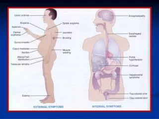
Physiologic changes seen in G.I. Parasitism • Loss of blood pale mucous membranes • diarrhea with loss of water and electrolyte disturbances • poor weight gains or even weight loss • protein loss
Physiologic changes seen in G.I. Parasitism • anorexia & reduced food intake • anemia • reduced digestion & absorption
Pathophysiology -Ostertagiasis • Life Cycle of Ostertagia • Adults mature in 17 to 21 days • Clinical Signs • diarrhea • loss of appetite • loss of weight or reduced weight gains
Pathophysiology -Ostertagiasis • Biochemical changes • in pH of abomasal contents • in levels of plasma pepsinogen • in serum proteins, particularly albumin - hypoalbuminemia
Pathophysiology -Ostertagiasis • Phases of Disease • Phase I,days 1-17,gastric glands invaded • Phase II,days 17 -35, adults emerge from the gastric glands • Phase III, > 35 days, recovery phase, adult expulsion
Pathophysiology -Ostertagiasis • There are two important types of cells in the gastric glands • Parietal cells - produce HCl • Chief cells - produce pepsinogen • The pepsinogen is activated by the HCl to pepsin - the functional hydrolytic enzyme of the abomasum
Pathophysiology -Ostertagiasis • The integrity of the epithelium is maintained by an area of fusion of the lipo-protein layers of the plasma membranes of the adjacent cells. This area of fusion is called the Zona Occludens
Phase I • Development of larvae in the gastric glands • This is where all the changes occur as the larvae develop and moult twice to become immature adults
Phase I • The inflammatory response induced by the growing larvae produce a constant erosion of the epithelial lining of the gastric glands • These cells are replaced by immature non-secretory epithelial cells
Phase I • At post mortem these parasitized glands can be seen as white raised nodules on the surface of the abomasum
Phase I • Abomasal pH remains at 2.0 to 2.5 • No diarrhea, infected animals eat well and gain weight • Plasma pepsinogen levels rise slightly • Significant changes are about to occur
Phase II • Begins about 17 days after infection • with the emergence of the young adults from the gastric glands • this further stretches and erodes the glands • the worms are large enough that they begin to destroy the surrounding cells • net effect is widespread erosion, destroying many functional zymogen and parietal cells
Phase II • The nodular effect in the abomasal mucosa is now widespread • The Moroccan leather appearance
Phase IIThree changes • An increase in the abomasal pH • directly attributable to the loss of parietal cells • pH goes from ~ 2 to ~ 7
Phase IISecond change • Reduced pepsinogen output as a result of loss of zymogen cells
Phase IIThird change • Enhanced permeability of abomasal mucosa, caused by: • failure to convert pepsinogen to pepsin • there is little conversion above pH 5 • failure to denature proteins in preparation for digestion • loss of bacteriostatic effect of low pH
Enhanced permeability of the mucosa results from the normal secretory epithelium being replaced by rapidly dividing immature cells whose tight junctions (zona occludens) are not fully formed • Thus the integrity of the stomach lining is lost and molecules can cross in both directions
Phase II • Pepsinogen will pass into the circulation from the lumen • thus resulting in high plasma pepsinogen levels • Plasma proteins, primarily albumin, will be lost into the gut • All via these leaky junctions
Phase II • Clinical Consequences • impaired digestion • loss of appetite • diarrhea • dehydration • weight loss or poor weight gains
Phase II • Impaired digestion • due primarily to the loss of pepsin activity • Loss of appetite • related to numbers • stimulate production of cholecystokinin • CCK depress appetite • parasites produce a substance that induces inappetence
Phase II • Diarrhea and Dehydration • reported for all g.i. Nematodes, except Haemonchus • follows the increased pH and in number of bacteria • there are usually high levels of osmotically actives substances (bacteria, undigested protein) in the gut • cause water to move into the intestine
Phase II • Weight loss • up to a 20% weight loss is possible • this may be due to: • loss of protein through leaky mucosa • anorexia • impaired digestion • this means that energy and protein are not coming from food intake and digestion, but from mobilization of protein reserves in the body, i.e. muscle
Phase III • Recovery can come about by: • removing the animals from pasture, from the source of infection • removing the worms with an anthelmintic • Within a relatively short period of time the differentiated status of the mucosa has returned • clinical signs subside and physiology returns to normal
Haemonchosis in SheepMost common observations • Unexpected deaths • Weakness • Anemia • Hypoproteinemia • Subcutaneous edema • Poor weight gains or weight loss