Download
1 / 6
60 likes | 82 Views
Direct Plagiarism

E N D
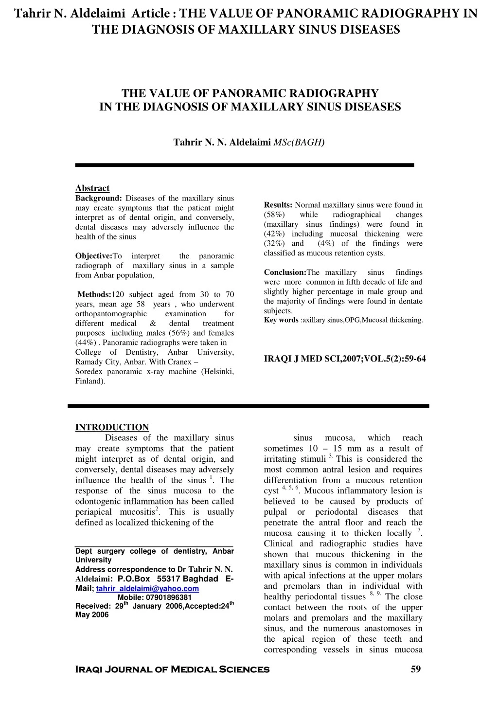
Tahrir N. Aldelaimi Article : THE VALUE OF PANORAMIC RADIOGRAPHY IN THE DIAGNOSIS OF MAXILLARY SINUS DISEASES THE VALUE OF PANORAMIC RADIOGRAPHY IN THE DIAGNOSIS OF MAXILLARY SINUS DISEASES Tahrir N. N. Aldelaimi MSc(BAGH) Abstract Background: Diseases of the maxillary sinus may create symptoms that the patient might interpret as of dental origin, and conversely, dental diseases may adversely influence the health of the sinus Results: Normal maxillary sinus were found in (58%) while radiographical (maxillary sinus findings) were found in (42%) including mucosal thickening were (32%) and (4%) of the findings were classified as mucous retention cysts. changes Objective:To radiograph of from Anbar population, interpret maxillary sinus in a sample the panoramic Conclusion:The maxillary were more common in fifth decade of life and slightly higher percentage in male group and the majority of findings were found in dentate subjects. Key words :axillary sinus,OPG,Mucosal thickening. sinus findings Methods:120 subject aged from 30 to 70 years, mean age 58 years , who underwent orthopantomographic different medical & purposes including males (56%) and females (44%) . Panoramic radiographs were taken in College of Dentistry, Ramady City, Anbar. With Cranex – Soredex panoramic x-ray machine (Helsinki, Finland). examination dental for treatment Anbar University, IRAQI J MED SCI,2007;VOL.5(2):59-64 INTRODUCTION Diseases of the maxillary sinus may create symptoms that the patient might interpret as of dental origin, and conversely, dental diseases may adversely influence the health of the sinus response of the sinus mucosa to the odontogenic inflammation has been called periapical mucositis2. This is usually defined as localized thickening of the sinus mucosa, which reach sometimes 10 – 15 mm as a result of irritating stimuli3.This is considered the most common antral lesion and requires differentiation from a mucous retention cyst believed to be caused by products of pulpal or periodontal penetrate the antral floor and reach the mucosa causing it to thicken locally Clinical and radiographic studies have shown that mucous thickening in the maxillary sinus is common in individuals with apical infections at the upper molars and premolars than in individual with healthy periodontal tissues contact between the roots of the upper molars and premolars and the maxillary sinus, and the numerous anastomoses in the apical region of these teeth and corresponding vessels in sinus mucosa 1. The 4, 5, 6. Mucous inflammatory lesion is diseases that 7. Dept surgery college of dentistry, Anbar University Address correspondence to Dr Tahrir N. N. Aldelaimi: P.O.Box 55317 Baghdad E- Mail; tahrir_aldelaimi@yahoo.com Mobile: 07901896381 Received: 29thJanuary 2006,Accepted:24th May 2006 8, 9.The close 59 Iraqi Journal of Medical Sciences Iraqi Journal of Medical Sciences
Tahrir N. Aldelaimi Article : THE VALUE OF PANORAMIC RADIOGRAPHY IN THE DIAGNOSIS OF MAXILLARY SINUS DISEASES PANORAMIC RADIOGRAPHY……… Tahrir Aldelaimi have been found to permit the spread of odonogenic pathological processes from the periodontium and pulpal spaces both directly and via vessels to the maxillary sinus both dentate and edentulous subjects. Prevalence figures ranging from 2% to 13% have been reported3, 12, 13. The diffuse mucosal thickening is more common with frequencies up to 50% of the radiographic incidental findings are included in the paranasal sinuses are more common in the maxillary sinus Bjorn et al.16and Lindhall et al. radiographic signs mucosal changes in the maxillary sinus in 10.6% of statistical sample of a Swedish population. Prevalence figures for sinusitis due to dental causes vary between 4.6 and 47%. However it has been suggested that, mucous retention cysts are insignificant clinically and only of radiograph interest 18. Further more mucosal thickening usually cause no symptoms, but occasionally they have been related to a variety of symptoms, mainly, facial pain, toothache3. 19. Mucosal thickening resolve when their caused symptomatic cases, removal of the cyst may be indicated (20,21,22). Myall et al in 1974 benign mucosal cyst is the most maxillarymolars Halstead in 1973 retention cysts are domeshape shadow of uniform density within the maxillary sinus whose base is continuous with the floor or the wall of the maxillary sinus and the free surface of the lesion should be smooth and sharply defined and adjacent to an air shadow. Also, there should be no osseous cortex Layon panoramic radiography in the diagnosis of maxillary antral pathosis. The disadvantage of panoramic radiography arises from their technique, distortion levels may reach 30% in the third molar region maxillary sinus is clearly imaged in panoramic radiography, but small changes out side the 2 –3 mm thick sharply depicted layer are not visualized in the normal panoramic projection, the roof of the maxillary sinus is not imaged because of superimposition of bones mucous cysts and thickening are usually well demonstrated as they almost always arises from the antral floor not from roof30, 29, 28, 23. Statistical analysis: includes percentages, mean, standard deviation and student "t" test. The finding was considered as statistically significant if the p value <0.005, Karl –person correlation (r) was used to find inter observer reliability (-1<r<+1). 27. However other mucosal 10, 11. In radiographical studies of 14. Mucous cysts which coffiecient of 15. 17found standing MATERIALS AND METHODS: 120 subject aged from 30 to 70 years, mean age 58±8 years, who underwent orthopantomographic examination for different medical & dental treatment purposes including 66 males (56%) and 54 females (44%). Panoramic radiographs were taken in college of Dentistry, Anbar University, Ramady City, Anbar. With Cranex – Soredex panoramic x-ray machine (Helsinki, Finland), All patient were referred to college of dentistry requesting OPG examinations, panoramic films were processed by Kodak automatic processor. The radiographs then were studied under standardized condition by two examiners (double blind technique) with the use of magnifying lens of radiographic viewer. radiographs were interpreted for these findings using radiographic criterion thickening and mucous retention cyst of the maxillary sinus (24,91,6). The mucous retention cyst is a well defined dome-shaped opacity outline arising from the floor of the maxillary sinus, while the mucosal thickening is represented by the more diffuse opacities along the margins of the sinus without well-defined rounded outline, as mentioned both are usually well demonstrated as they almost always arises from the antral floor not from roof.(30,29,28,23). of long mucous cysts and headache and is however removed. In surgical RP X-omat 6stated that independent .Its incidence varies by 20To 9.6% in one round, ovoid or Panoramic a standardized of mucosal 6. 24has discussed the reliability of with convex main dynamic projection 25, 26. The 60 Iraqi Journal of Medical Sciences Iraqi Journal of Medical Sciences
Tahrir N. Aldelaimi Article : THE VALUE OF PANORAMIC RADIOGRAPHY IN THE DIAGNOSIS OF MAXILLARY SINUS DISEASES PANORAMIC RADIOGRAPHY……… Tahrir Aldelaimi Result The study sample was representing (2%) (Table 3). Regarding the sex (Table 4), findings were slightly higher in the males rather than the females, where the mucosal thickening was found in (18%) within gender. mucous retention cysts were found in (3%) within the gender. Table 5 showed that the prevalence of mucosal thickening in dentate and edentulous patients representing (20%) and (12%). Other maxillary sinus findings were also recorded in this study. There were (4%) of patients showed impaction & displacement of a tooth inside the maxillary sinus. maxillary teeth or tooth were either canine or second molar, also severe pneumaitization of the maxillary sinus floor down to the alveolar crest was seen in (2%). including 66 (56%) males and 54 (44%) females with age ranged from 30-70 years of mean age 58±8 year. The distribution of the number of patients and age summarized in (Table 1). Normal radiographical (maxillary findings) were 70 subjects (58%) while maxillary sinus findings were found in 50 subjects (42%). Including mucosal thickening in 38 patients (32%) and 4 patients (4%) have mucous retention cysts (Table 2). The highest percentage of mucosal thickening was found that in the age group represent (14%) within the age group. Regarding the mucous retention cyst the highest percentage was found also among the age group years the maxillary groups are While the sinus (40-49) years The impacted TABLE 1: T he distribution of age group in relation to sex AGE GROUP 30-39 40-49 50-59 60-69 TOTAL 120(100%) MALE % ٥ 24% 18% 9% 66(56%) FEMALE % ٣ 28% 9% 4% 54(44%) TABLE 2: The distribution of radiographical maxillary sinus findings* MAXILLARY SINUS FINDINGS Normal Mucosal thickenings Mucous retention cyst Others PERCENT 70(58%) 38(32%) 4(4%) 4(4%) 3(2%) Root inside antrum Pneumatization (sinus floor to alveolar ridge) TOTAL 120 (100)% * r=0.9 61 Iraqi Journal of Medical Sciences Iraqi Journal of Medical Sciences
Tahrir N. Aldelaimi Article : THE VALUE OF PANORAMIC RADIOGRAPHY IN THE DIAGNOSIS OF MAXILLARY SINUS DISEASES PANORAMIC RADIOGRAPHY……… Tahrir Aldelaimi TABLE 3: The distribution of maxillary sinus finding in relation to patients age group AGE GROUP NORMAL MUCOSAL THICKENING MUCOUS RETENTION CYST 0% 2% 1% 1% 4(4%) OTHERS 30-39 40-49 50-59 60-69 TOTAL 120(100%) 4% 26% 17% 11% 70(58%) 2% 14% 10% 6% 38(32%) 1% 2% 2% 1% 7(6%) TABLE 4: The distribution of radiographical maxillary sinus findings in relation to sex SEX NORMAL MUCOSAL THICKENING MUCOUS RETENTION CYST 3% 1% 4(4%) OTHERS MALE FEMALE TOTAL 120(100%) 32% 26% 70(58%) 18% 14% 38(32%) 4% 2% 7(6%) TABLE 5: The distribution of radiographical maxillary sinus finding in relation to maxillary arch MAXILLARY ARCH NORMAL MUCOSAL THICKENING MUCOUS RETENTION CYST 2% 2% 4(4%) OTHERS DENTATE EDENTULOUS TOTAL 120(100%) 34% 24% 70(58%) 20% 12% 38(32%) 3% 3% 7(6%) DISCUSSION The prevalence of mucous and diffuse mucosal thickening in all the paranasal sinuses been as high as radiographs taken for indications other than suspected sinus disease magnetic resonance imaging study of incidental findings in the paranasal sinuses of 438 subjects, the prevalence of incidental findings in all sinuses was 37.5% and they were most common in the maxillary sinus of the maxillary sinus findings among elderly edentulous in previous studies of variable ranges, however figures ranging from 2.6% to 20% have been reported10,12. In a study of Soikkonen and Ainomo in 199414, The prevalence of mucous cysts and diffuse mucosal thickening in the maxillary sinuses of elderly edentulous subject was 7% studies of rounded shadows (mucous cysts) in maxillary sinus found in both dentate and edentulous subject with figures ranging from 2% to 13%12, 3, 13. has 50% occasionally in facial 32. In 32. The prevalence 62 Iraqi Journal of Medical Sciences Iraqi Journal of Medical Sciences
Tahrir N. Aldelaimi Article : THE VALUE OF PANORAMIC RADIOGRAPHY IN THE DIAGNOSIS OF MAXILLARY SINUS DISEASES PANORAMIC RADIOGRAPHY……… Tahrir Aldelaimi Our figures of 4% for the prevalence of mucous retention within that range. According to mattila,29the prevalence of mucous cysts is not age-dependant. This was in accordance with our results, where no statically significant difference was found between age groups (p<0.005). In studies including younger age groups, maxillary sinus findings have been most prevalent in the third decade and they have also been found to be more prevalent in men contrary with ours, where the findings were more common in the fifth decade of life and comes in accordance with ours regarding the slightly higher percentage in the male group. In the rather wide age-range of the present study old subjects, the number of maxillary sinus findings showed no age-dependent tendencies. The diffuse mucosal thickening, however, were more prevalent in the younger age group, the majority of the diffuse mucosal thickening were found in dentate subject of younger age group. More important (than dental origin) is that allergic sinusitis especially due to dust inhalation especially in this region of Iraq due characteristic of the region and it can be suspected that odontogenic causes may not be a major contributing factor in their formation. This result comes in accordance with previous who stated that, the prevalence maxillary sinus findings in sites of periapical or periodontal pathosis without pathologic findings have also been similar nor ours support the findings of Halstead in 197320, Who reported that a possible odontognic cause could be indicated in 90% of subjects with maxillary sinus findings. Regarding The diffuse mucosal thickenings, it was reported that those findings always indicate the presence stimuli, after an infection of dental origin Although showed difference (p<0.005) between dentate and edentulous patients in relation to the mucosal thickening found in the floor of the sinus. It has been stated that, the chronic apical periodontitis, deep infra-bony pockets are usually unaccompanied by subjective symptoms. Their accurate diagnosis may sometimes be vital to the patient, for if the host resistance for same reason, it will give this infection the opportunity to become exacerbated and cause acute sinusitis, whereas the possibility also exists of further spread systemic manifestation34, 28. no statistical significant any major 12, 3, 27. This result is on the REFERENCES 1. Margot VD and Dale AM. Disorders of the maxillary sinus. Dental America.1994; 38:1-8. 2. Langland OE, Langlais RP, Mcdard WD. Panoramic radiography, Philadelphia, lea&febiger; 1989.pp 406. 3. Gooz P and White S. Oral radiology: principles and interpretation. 3RdEd. St louis. Mosby, 1994:pp 602-610. 4. Gardner DG. Pseudocysts And Retention cysts of the maxillary sinus. Oral Surg. Oral Med Oral Path.1984; 58: 561-567. 5. Gardner DG, Gullame PJ. Mucoceles of maxillary sinus. Oral Surg Oral Med Oral Path .1986; 62: 538-543. 6. Myall RW, Eastephan PP and Silver IG. Mucous retention cyst of the antrum.JADA.1974; 89: 1338-1342. 7. Worth HM and Stonman DW. Radiographic interpretation of antral mucosal change due to localized dental infection. Assoc.1972; 38: 111-115. 8. Connor SE, Chavda SV and Pahor AL. Computed Tomographic evidence of dental Restoration aetiological Factor for maxillary sinusitis. J laryngolotol.2000; 114:510-513. 9. Falk H, Ercson S effect of periodontal treatment on mucous membrane bin the maxillary sinus. J Clinc Periodontal.1986; 13: 217-222. 10. Killy HC, Kay sinus&dental implication, 3rd edition, wright. 1981: pp10-23. 11. Ohba T and Katayama H. Comparison of panoramic radiography and Water’S Projection in the diagnosis of maxillary sinus diseases. Oral Surg Oral Med Oral Path .1976; 42: 534-538. 12. Allard RH, Vander Wl. Mucousal antral cysts. Oral Surg Oral Med Oral Path. 1981; 51: 2-9. clinic Of North 2nd edition, to sentimental J Can Dent and in sites and Hugoson A. The 33. Neither that findings LW. The maxillary of irritating 8,14. our results 63 Iraqi Journal of Medical Sciences Iraqi Journal of Medical Sciences
Tahrir N. Aldelaimi Article : THE VALUE OF PANORAMIC RADIOGRAPHY IN THE DIAGNOSIS OF MAXILLARY SINUS DISEASES PANORAMIC RADIOGRAPHY……… Tahrir Aldelaimi 13. Wright Maxillary sinus. Laryngoscope.1946; 56: 455- 456. 14. Soikkonen K, Ainamo O and Wolf J. Radiographic findings in the jaws of clinically edentulous old people living at home in Helsinki Finland. Acta Odontol Scand.1994; 52: 229-233. 15. Cooke ID, Hadlley DM. MRI of paranasal sinuses: incidental abnormalities relationship to symptoms. J laryngeolotol. 1991; 105: 278-281 16. Bjorn H, Holmberg K and Nylander G. Maxillary sinus In Odontolog.1967; 18:83-114. 17. Lindhall IL, Melen J, Ekedal C and Holm S. Chronic maxillary Otolaryngologica.1982; 93: 147-150. 18. Killey HC and Kay IW. Benign mucosal cysts of the maxillary sinus. Int. Surg. 1970; 53: 235-238. 19. Rhodus NI. The prevalence and clinical significance of maxillary sinus mucous retention cysts in a general clinic population. Ear nose throat.1990; 69: 82-87. 20. Salstead CL. Mucosal cysts of the maxillary sinus. JADA 1973; 87:14-20. 21. Fisher EW: Round shadows in the maxillary sinus. Laryngoscope.1946; 56: 455-456. 22. Millhon JA and Brown HA. Cysts Arising From The Mucosa of the maxillary sinus as seen in dental roentgenogram. Am J Orthod. 1944; 30: 12-14. 23. Paparella MM. Mucosal Maxillary sinus. Arch Otolaryngol.1963; 77: 650-652. 24. Layon HE. Reliability radiography in the diagnosis of maxillary sinus pathosis. Oral Surg.1973; 35: 124-126. 25. Christen AG and Segreto VA. Distortion And Artifacts encountered Radiography. JADA.1968; 77: 109-110. 26. Kite OW. Radiation And Image distortion in the panorex x-ray unit. Oral Surg. 1962; 15: 1201-1205. 27. Mcgowan D, Baxter P and James J. The maxillary sinus and its dental implication. Oxford. 28. Wright.1993,pp 1-153. 29. Naschitz JE and Yeshurun D. Occult infection in the facial area presenting as fever of unknown origin. Isr Med sci. 1985; 21: 995-998. 30. Mattila K. Roentgenologicl investigation into the between Periapical lesions And condition of the mucous membrane of maxillary sinus. ActA Odont Scand.1965; 23: 1-19. 31. Kwapis BJ. Whitten JP:Mucosal cysts of the maxillary sinus. J Oral Surg. 1971; 29:561-566. 32. Wilson PS and Grocutt M. Thickening on sinus x-ray and laryngolotol.1996; 104: 694-695. 33. Ohba T. Value and limitation of panoramic radiography in the diagnosis of maxillary sinus pathosis. Int. J. Oral Surg. 1977; 6: 211-214. 34. McDonalds and Kawasaki DS. Mucosal antral cysts in a Dentomaxillofac. Radiol.1993; 22: 208-210. 35. Huebner GR and Groat D. The role Of Dental disease in fever of unknown origin. Postgrad. Med. 1986; 79: 275-278. RW. Round shadows In The Chinese population. and their Periodontal disease. sinusitis. Acta cysts of the of panoramic In Panorex its significance: J 64 Iraqi Journal of Medical Sciences Iraqi Journal of Medical Sciences View publication stats View publication stats