Download
1 / 16
200 likes | 2.74k Views
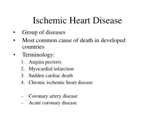
Ischemic Heart Disease. Dr. Fletcher. Week 3/ CVS module 9-11-2003. Ischemia heart disease. Restriction of blood supply, relative to demands of the heart. Ischein = to restrict. Recall: Heart has high : - Work load. - Metabolic Rate. - Aerobic metabolism.

E N D
Ischemic Heart Disease Dr. Fletcher. Week 3/ CVS module 9-11-2003
Ischemia heart disease • Restriction of blood supply, relative to demands of the heart. • Ischein = to restrict. Recall: • Heart has high : - Work load. - Metabolic Rate. - Aerobic metabolism. - Oxygen demands.
Features of Ischemia heart disease : • Coronary Obstruction. • Cardiac Pain. • Cardiac Ischemia lesions.
1) Coronary Obstruction: • Obstruction of coronary arteries decrease blood to heart.
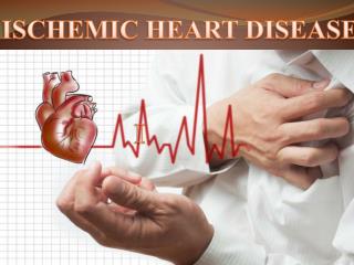
2) Cardiac Pain: Common Characteristics : • Sever pain gripping, crushing, arresting. • Chest “Retrosternal”, behind the sternum • Radiating to left arm, neck and jaw.
Coronary obstruction/Cardiac pain/Cardiac Ischemia lesion Obstruction: Impediment. Stenosis Narrowing of blood vessle Pain : Angina Pectoris Cardiac lesions Ischemia fibrosis. Narrow lumen I) Obstruction II) Occlusion Cardiac lesions Infarct (necrosis). Pain : Infarct Pain Occlusion: Closed vessel Closure of the lumen
I) Stenosis: • It is narrowing without complete closure this is a gradual process. • As a result of slow gradual changes it will give chance for adaptation by forming anastomosis ( other area feed the Ischemic area) Partial Ischemia. • Lesions: Occasional cell atrophy. It is not a vacuum, because fibrous tissue replace the space ( Fibrosis intravenous).
Pain Angina Pectoris: • General chest pain. • Its specific characteristic are: 1) Induced by exertion. 2)Paroxysmal (~ 15 minutes) (to differentiate between it and other chest pain). 3) Relieved by rest. 4) Relieved by Nitroglycerin vasodilatations.
II) Occlusion: • Sudden onset ( due to thrombosis, plaque rupture) • Complete Ischemia to the area of heart. • Give area of infarction sizable.
Pain of infarct type: (Important for distinguishing) 1) Very sever. 2) Prolonged pain - at least 30 min (not 15 min) - often 1 hours. - Some cases 6 hrs ( have to be controlled byopiates). 3) Not induced by exertion. 4) Not relieved by rest. 5) Not relieved by nitroglycerin.
3) Cardiac Lesions: • Ventricle lesions ( 97%). • Right ventricle lesion extensions in the left or septal.
1. Functional disruption of the heart: Arrhythmia due to Ischemia ( local) of conducting system. it can be picked up by ECG & might end up with sudden death.
2. Diffuse ischemic fibrosis: • It is Patchy muscle fiber atrophy • There is a replacement fibrosis ( strengthens). • Post Mortem examination: We see – Grayish-white sheen. - ill defined Vague
3. Infarction: due to Occlusion • Post mortem Features : • Inapparent until >18 hrs survival. • They become Apparent after 24 hrs. • Infarct site are Yellow due to Autolysis at 37C (viable muscle). • When tissue necrosis occur • The tissue release many substances such as LDH, CPK and Troponin • Induce inflammatory reaction migration of neutrophils to the site of necrosis and Increase phagocytosis. • Electrolyte imbalance (increase Na and decrease K)
Sudden Death: Causes: 1) Arrhythmia Ventricular fibrillation. 2) Ischemia : a) re-entrant circuit b) damaged conducting system, coronary lesions (hard to find). 3) Massive Acute infarct: infarct is not visible but coronary thrombosis easily found.