Download
1 / 70
710 likes | 1.7k Views
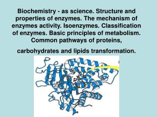
Proteins: Their Structure and Biological Functions. Biological Functions of Proteins. Proteins are the agents of biological function Enzymes - Ribonuclease Regulatory proteins - Insulin, PCNA Transport proteins - Hemoglobin Structural proteins - Collagen

E N D
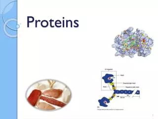
Biological Functions of Proteins Proteins are the agents of biological function • Enzymes - Ribonuclease • Regulatory proteins - Insulin, PCNA • Transport proteins - Hemoglobin • Structural proteins - Collagen • Contractile proteins - Actin, Myosin • Protective proteins - Antifreeze proteins
Protein structure often provides clues about protein function Unrelated proteins assume similar structures to fulfill common functions
Peptides • Short polymers of amino acids • Each unit is called a residue • 2 residues - dipeptide • 3 residues -tripeptide • 12-20 residues - oligopeptide • many - polypeptide
Protein One or more polypeptide chains • One polypeptide chain - a monomeric protein • More than one - multimeric protein • Homomultimer - one kind of chain • Heteromultimer - two or more different chains • Hemoglobin, for example, is a heterotetramer; it has two alpha chains and two beta chains
Proteins - Large and Small • Insulin - A chain of 21 residues, B chain of 30 residues -total mol. wt. of 5,733 • Glutamine synthetase - 12 subunits of 468 residues each - total mol. wt. of 600,000 • Connectin proteins - alpha - MW 2.8 million! • beta connectin - MW of 2.1 million, with a length of 1000 nm -it can stretch to 3000 nm!
Amino acid composition provides some (limited) clues about protein structure-function
The Sequence of Amino Acids in a Protein • is a unique characteristic of every protein • is encoded by the nucleotide sequence of DNA • is thus a form of genetic information • is read from the amino terminus to the carboxyl terminus
The levels of protein structure - Primary sequence - Secondary local structures - Tertiary overall 3-dimensional shape - Quaternary subunit organization
What forces determine the structure? • Primary structure - determined by covalent bonds • Secondary, Tertiary, Quaternary structures - all determined by weak forces
The Role of the Sequence in Protein Structure All of the information necessary for folding the peptide chain into its "native” structure is contained in the primary amino acid structure of the peptide.
Sequence Determination Frederick Sanger was the first - in 1953, he sequenced the two chains of insulin. • Sanger's results established that all of the molecules of a given protein have the same sequence • Proteins can be sequenced in two ways: - real amino acid sequencing - sequencing the corresponding DNA in the gene
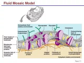
Nature of Protein Sequences • Sequences and composition reflect the function of the protein: • Membrane proteins have stretches of hydrophobic residues, whereas fibrous proteins may have atypical sequences • Homologous proteins from different organisms have similar sequences e.g., cytochrome c is highly conserved
Phylogeny of Cytochrome c • The number of amino acid differences between two cytochrome c sequences is proportional to the phylogenetic difference between the species from which they are derived • This observation can be used to build phylogenetic trees of proteins • This is the basis for studies of molecular evolution
The Coplanar Nature of the Peptide Bond Six atoms of the peptide group lie in a plane
The Peptide Bond • is usually found in the trans conformation • has partial (40%) double bond character • is about 0.133 nm long - shorter than a typical single bond but longer than a double bond • Due to the double bond character, the six atoms of the peptide bond group are always planar. • N partially positive; O partially negative
Secondary Structure The atoms of the peptide bond lie in a plane • The resonance stabilization energy of the planar structure is 88 kJ/mol • A twist about the C-N bond involves a twist energy of 88 kJ/mol times the square of the twist angle. • Twists can occur about either of the bonds linking the alpha carbon to the other atoms of the peptide backbone
Consequences of the Amide Plane Two degrees of freedom per residue for the peptide chain • Angle about the C(alpha)-N bond is denoted phi • Angle about the C(alpha)-C bond is denoted psi • The entire path of the peptide backbone is known if all phi and psi angles are specified • Some values of phi and psi are more likely than others.
Steric Constraints on phi & psi Unfavorable overlap precludes some combinations of phi and psi • phi = 0, psi = 180 is unfavorable • phi = 180, psi = 0 is unfavorable • phi = 0, psi = 0 is unfavorable
Classes of Secondary Structure All these are local structures that are stabilized by hydrogen bonds • Alpha helix • Beta sheet (composed of "beta strands") • Tight turns (aka beta turns or beta bends)
The Alpha Helix • First proposed by Linus Pauling and Robert Corey in 1951 • A ubiquitous component of proteins • Stabilized by H-bonds
The Alpha Helix • Residues per turn: 3.6 • Rise per residue: 1.5 Angstroms • Rise per turn (pitch): 3.6 x 1.5A = 5.4 Angstroms • The backbone loop that is closed by any H-bond in an alpha helix contains 13 atoms • phi = -60 degrees, psi = -45 degrees
The Beta-Pleated Sheet Composed of beta strands • Also first postulated by Pauling and Corey, 1951 • Strands may be parallel or antiparallel
The Beta Turn (aka beta bend, tight turn) • allows the peptide chain to reverse direction • carbonyl C of one residue is H-bonded to the amide proton of a residue three residues away • proline and glycine are prevalent in beta turns
Steric Constraints on phi & psi • G. N. Ramachandran was the first to demonstrate the convenience of plotting phi,psi combinations from known protein structures • The sterically favorable combinations are the basis for preferred secondary structures
Predictive Algorithms If the sequence holds the secrets of folding, can we figure it out?
Tertiary StructureSeveral important principles: • The backbone links between elements of secondary structure are usually short and direct • Proteins fold to make the most stable structures (make H-bonds and minimize solvent contact
Tertiary Structure So, how do proteins fold?
Weak Forces are Responsible for Protein Folding What are they? What are the relevant numbers? • van der Waals: 0.4 - 4 kJ/mol • hydrogen bonds: 12-30 kJ/mol • ionic bonds: 20 kJ/mol • hydrophobic interactions: <40 kJ/mol