Download
1 / 1

E N D
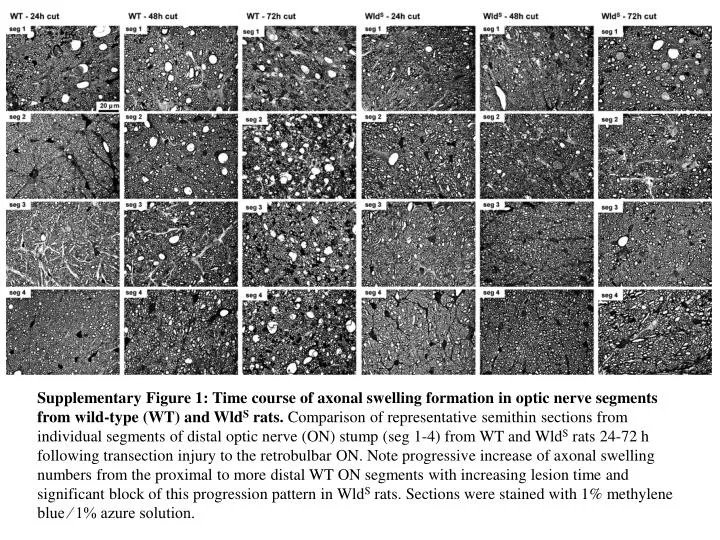
Supplementary Figure 1: Time course of axonal swelling formation in optic nerve segments from wild-type (WT) and WldS rats. Comparison of representative semithin sections from individual segments of distal optic nerve (ON) stump (seg 1-4) from WT and WldS rats 24-72 h following transection injury to the retrobulbar ON. Note progressive increase of axonal swelling numbers from the proximal to more distal WT ON segments with increasing lesion time and significant block of this progression pattern in WldS rats. Sections were stained with 1% methylene blue ⁄ 1% azure solution.