Download
1 / 13
150 likes | 435 Views
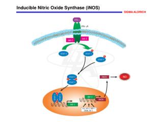
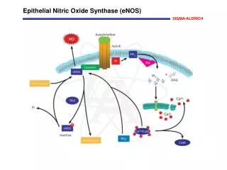
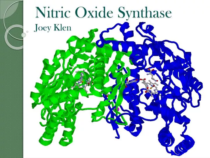
Nitric Oxide Synthase Joey Klen. Introduction. Nitric oxide (NO) is an important signaling biomolecule and a cytotoxin. NO is produced by nitric oxide synthase (NOS) Neuronal ( nNOS ) Neuronal tissues; NO as a neurotransmitter Inducible ( iNOS ) Macrophages; NO as a cytotoxin

E N D
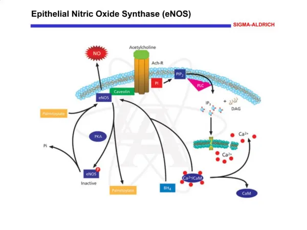
Introduction • Nitric oxide (NO) is an important signaling biomolecule and a cytotoxin. • NO is produced by nitric oxide synthase (NOS) • Neuronal (nNOS) • Neuronal tissues; NO as a neurotransmitter • Inducible (iNOS) • Macrophages; NO as a cytotoxin • Endothelial (eNOS) • Endothelial cells; NO as a vasodilator
Reaction • Catalizes a 2-step reaction from L-Arg to L-citrulline and NO • Two major domains: • Catalytic (oxygenase) domain: • Heme • L-Arginine • Tetrahydrobiopterin (H4B) • Reductase Domain • NADPH • FAD, FMN
Step 1 of Mechanism • Conversion of L-Arg to Nw-hydroxy-L-arginine (NHA) • Similar to cytochrome P450 NADPH H4B
Step 2 of Mechanism • Conversion of NHA to L-citrulline and NO • Not like P450 NADPH H4B
Heme Binding • Cys-415 binds to heme Fe • Tyr-706 binds to heme propionate oxygen • Trp-409 can p-stack with heme ring
L-Arginine/NHA Binding • Glu-592 binds in three places • Pro-565 H-bonds to a H2O, which then binds to the guanidinium N of L-arg. • Hydroxylated N-H of NHA binds to O2 oxygen bound to heme Fe
H4B Binding • Carbonyl H-Bonds • Ser-334 • Trp-678 • Phe-691 • Arg-596 • Heme propionate group H4B
Zinc Tetrathiolate Cluster • NOS is only active as a homodimer; the monomeric form is inactive • The dimer is held together by a Zn2+ ion complexed to 2 pairs of Cys residues from each paired monomer • High [NO] can cause S-nitrosation of 2 Cys residues, which leads to release of Zn and formation of inactive NOS monomers
Sequence Alignment Key: Alpha Helices Beta Sheets Heme Binding Residues L-Arg/NHA Binding Residues H4B Binding Residues Zn Tetrathiolate Cys Residues
Kinetic Data A. Rate of NO production of WT rat nNOS monitored by Hb assay at room temperature. B. Lineweaver-Burk plot for dtermination of Km and Vmax.
References • (Structure Paper) Doukov, T., Li, H., Soltis, M., and Poulos, T. L. (2009) Single crystal structure and absorption spectral characterizations of nitric oxide synthase complexed with Nw-hydroxy-L-arginine and diatomic ligands. Biochemistry 48, 10246-10254. • Kerwin, J. R. Jr., Lancaster, J. R. Jr., and Feldman, P. L. (1995) Nitric oxide: a new paradigm for second messengers. J. Med. Chem. 38, 4343-4362. • Mitchell, D. A., Erwin, P. A., Michel, T., and Marletta, M. A. (2005) S-Nitrosation and regulation of inducible nitric oxide synthase. Biochemistry 44, 4636-4647. • Raman, C. S., Li, H., Martasek, P., Kral, V., Masters, B. S. S., and Poulos, T. L. (1998) Crystal structure of constitutive endothelial nitric oxide synthase: a paradigm for pterin function involving a novel metal center. Cell 95, 939-950. • Li, D., Kabir, M., Stuehr, D. J., Rousseau, D. L, and Yeh, S. R. (2007) Substrate- and isoform-specific dioxygen complexes of nitric oxide synthase. J. Am. Chem. Soc. 129, 6943-6951. • Fang, J., Ji, H., Lawton, G. R., Xue, F., Roman, L. J., and Silverman, R. B. (2009) L337H Mutant of rat neuronal nitric oxide synthase resembles human neuronal nitric oxide synthase toward inhibitors. J. Med. Chem. 52, 4533-4537