Download

1 / 31
360 likes | 698 Views
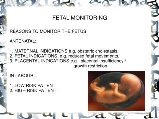
INTRAPARTUM FETAL MONITORING. Means careful vigilance of fetal behaviour during labour Aim of IP surveillance is the birth of a healthy baby in good condition with minimum intervention. Guidelines for methods and frequency IFM. Methods of fetal monitoring :

E N D
Means careful vigilance of fetal behaviour during labour Aim of IP surveillance is the birth of a healthy baby in good condition with minimum intervention.
Methods of fetal monitoring : • Intermittent FHR. Auscultation – stethoscope, fetoscopedoppler. • Biophysical - before and after amniotomy. • Electronic fetal monitoring • Fetal acoustic stimulation test • Fetal scalp stimulation • Umbilical Doppler • Intrapartum monitoring with fetal ECG wave form
Biochemical • Fetal blood sampling • Fetal pubeoximetry • Lactate level • Near infrared spectroscopy
Causes of Tachycardia : • Maternal / fetal infection • Maternal / fetal anemia • Fetal compromise • Drugs like adrenergic Ritodrine
Causes of bradycardia : • Fetal hypoxia / acidosis • Drugs – pethidine, local anesthelic, epidural, methyldopa, Mgso4, propranolol. • FH conduction defects (SLE)
Intermittent FHR monitoring : • Traditional • 30-45 seconds after a uterine contraction • Adequate for a low risk pregnancy • Monitored every 15 min. in 1st stage and every 5 min. in 2nd stage.
Limitations : • Transient significant abnormality in between observation is overlooked. • Inherent human error. • Difficult in cases of obesity / polyhydramnios.
Meconium in Liquor : • Meconium staining of liquor after ROM gives a crude idea of intrauterine jeopardy. • Absent in premature body, imperforated anus, Rh incompatibility. • Meconium in liquor in a potential sign of hypoxia. • Hypoxia – increased vagal response – increased peristaltic activity • Relaxation of anal sphincter – passage of M. • Presence of meconium and non reassuring FHR. • Patten requires intervention.
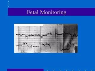
Continuous FHR monitoring : • Allow for the assessment of sequential information regarding fetal condition. • FHR is detected by placing a US transducer on maternal abdomen wall. • Coupling gel is applied between the transducer and abdomen wall and the transducer is held in position by a belt.
Continuous electronic fetal monitoring : • High risk group • External • Internal Advantages : • EFM improves perinatal mortality • Detect hypoxia early • Improvement of intrapartum death by 3 ford. • Important record for medico legal purpose.
Drawbacks : • Expensive • Training required for interpretation • Mother is confined to bed. • Cs rates are high.
Indications for EFM : • Antenatal : • Hypertensive disorder of pregnancy. • Pre-existing DM / GDM • APH • Maternal medical disorder • Maternal • Obesity • Fetal : • IUGR, PTL, Oligohydramnios. • Abnormal UAD, is immunization • MP, breech decreased.
Intrapartum EFM : • Vaginal bleeding in labour • Previous LSCS • PROM • Induced labour • Augmented labour • Post-term pregnancy • Meconium staining of AF • Abnormal FHR on auscultation.
Admission Test : • A short CTG for 20 minutes on admission in labor in low risk pregnancies. • Intermittent monitoring is recommended for normal Ad. Test. • When admission test is abnormal continuous CTG is recommended. • Identify a subgroup of fetuses that would be benefited from CEFM.
Decelerations Interpretation of an CTG : • Baseline FHR : 110-160 BPM • Baseline variability is the oscillation of FHR excluding the accelerations and decelerations. • Baseline of 5-25 BPM sign of fetal well being. • Reduced variability is seen in hypoxia, sleep phase cong. Malformation. • Drugs – sedatives, Mgso4 antihypertensives, mater acedmia.
Acceleration – increase in FHR by 15 BPM or > lasting for 15 seconds. • Decelerations : • Early – Head compression • Late • Variable – cord compression.
Prolonged deceleration last >120 sec. 90min) • Sinusoidal pattern - stable baseline FHR with fixed baseline variability without any acceleration – fetal anemia, FH, fetal hypoxia.
Classification of IP CTG • Normal trace / reassuring • Atypical / nonreassuring • Abnormal.
Decelerations : • Early : Occurs with uterine contractions and are due to fetal compression. Amplitude of D is rarely > 40 BPH. They are benign / or no particular significance. • Variable : They have erratic shape and appearances. Have variable relationship to uterine contraction. Due to U cord compression. Potentially dangerous to fetus. • Late : Occurs late in relation to uterus contraction. Represent fetal hypoxia.
Management of abnormal FHR patterns : • Move the mother to the lateral position • Correct maternal hypotension • Discontinue uterine stimulants • Tocolysis to correct hypertones • Administer oxygen 6-8 L • Vaginal examination, perform ARM • Inform anaesthelist / nursing for emergency delivery • Inform pediatricians.
VAST : • Utilises an artificial larynx to stimulat6e fetus. • Scalp stimulation test • Fetal response is noted on CTG • Non hypoxic fetus will exhibit acceleration. • Improves the sensitivity of admission test.
Pulse oximetry : • Measures fetal oxyhaemoglobin saturation (below 40%). • Sensor is placed against the fetal check after ARM.
Lactate level measurement : • Serum lactate level is known indicator of anaerobic respiration. • Normal levels <2.8 mmol/L. • 2.9 – 3.08 mmol /L are suspicious > 3.08 mmol/L is abnormal.
Near infrared spectroscopy : • Method under evolving. • Light passed thro the detector placed on fetal head will be reflected back depending on the oxygenation and amount of blood flow.
Fetal acid – base measurements : • Persistent variable deceleration >45 min. • Late deceleration >30min. • Meconium SAF with non reassuring FHR Technique : • Patient in (L) lateral position. • PV • Fetal head is visualized thro’ the amnioscope – cleaned and silicone grease applied.
Ethyl chloride sprayed - hyperemia – with the help of blade 2mm (scalp) deep cut is made on scalp and then rotated thro’ 900. A drop of blood appears with is collected into capillary tube. • Normal PH • < 7.2 – acidosis • 7.2 – 7.24 preacidosi • > 7.25 – normal
C/I for fetal blood sampling : • Obvious indication for delivery • Face presentation • Maternal infections – HIV • Chorioamnionites • Prematurity • Fetal hemophilia.
INTERPRETATION : Baseline : 120-160 BPM – 110-150 BPM • Bradycardia < 120 BPM • Mild FHR 100-120 • Moderate 80-100 • Severe <80 • Tachycardia : > 160 BPM • Mild 161-180 • Severe > 180
Variability : 5-25 BPM • Increased variability is seen during fetal breathing and fetal body drug Mgso4. movements. • Decreased variability is seen in fetal compromise administration analgesic