Download
1 / 39
400 likes | 628 Views
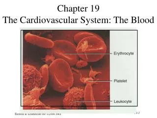
Chapter 10: Blood. Blood. The only fluid tissue in the body Classified as a connective tissue Notable: collagen & elastin absent from blood Dissolved fibrin proteins become evident during clotting Components of blood Living cells Formed elements – RBC & WBC Non-living matrix Plasma.

E N D
Blood • The only fluid tissue in the body • Classified as a connective tissue • Notable: collagen & elastin absent from blood • Dissolved fibrin proteins become evident during clotting • Components of blood • Living cells • Formed elements – RBC & WBC • Non-living matrix • Plasma
When Blood is centrifuged • Plasma rises to the top (55%) • Erythrocytes sink to the bottom (45%)5 • This is known as the hematocrit • Normal hematocrit: 47% +/- 5%: males • 42% +/-5%: females • Buffy coat contains leukocytes & platelets (<1%) • Thin, whitish layer that is between erythrocytes & plasma
Physical Characteristics of Blood • Sticky, opaque fluid – metallic taste • More dense than water, 5x more viscous • Colors: • Oxygen rich blood – scarlet red (bright) • Oxygen poor blood – dull red • pH is between 7.35 – 7.45 • Blood temperature is always slightly higher than body temperature – around 100.4F • In a healthy man, blood volume is about 5-6 liters or about 6 quarts • Women have less blood, about 3.5 – 5 liters • Blood makes up about 8% of body weight
Functions of Blood • All concerned with: • substance distribution • regulating blood levels of particularly substances • bodily protection • Distribution: • Oxygen to lungs • Nutrients to digestive system • Transporting metabolic wastes from elimination sites (lungs, kidneys, liver) • Hormones from glands to target organs
Functions of Blood • Regulation: • Maintain appropriate body temperature by absorbing and distributing heat through body, encouraging skin for heat loss • Maintain normal pH, blood holds an “alkaline” reserve to raise pH when necessary • Maintain adequate fluid volume in circulation • Salts & blood protein – prevent excess fluid loss • Protection • Prevents blood loss • Prevents infection
Blood Plasma • Straw colored, sticky liquid • Composed of approximately 90% water • Includes over 100 dissolved substances: • Nutrients • Salts (electrolytes) • Respiratory gases (CO2 & O2) • Hormones • Plasma proteins • Waste products • Uric acid, creatinine, lactic acid, ammonium salts
Blood Plasma: Plasma Proteins • Most abundant solutes in plasma • Made mostly by the liver • Proteins include: • Albumin: regulates osmotic pressure • The pressure that keeps water in the blood • Essentially carries all proteins around the blood • High albumin almost always caused by dehydration • Low albumin can come from liver disease, kidney disease, & malnutrition • Globulins: • Alpha, beta: transport proteins that bind to lipids, metal ions, fat-soluble vitamins • Gamma: antibodies released during an immune response • Clotting proteins: help to stop blood loss when a blood vessel is injured • Antibodies: help protect the body from pathogens (disease)
Blood Plasma • Acidosis: blood becomes too acidic • Liver failure, kidney failure • Alkalosis: blood becomes too basic • Main cause: hyperventilation – resulting in a loss of CO2 • Other causes: prolonged vomiting, Cushing’s syndrome, severe dehydration • In each scenario, the respiratory system & kidneys help restore blood pH to normal
Formed Elements • Erythrocytes • Red blood cells (RBCs) • Leukocytes • Lymphocytes • Both are white blood cells (WBCs) • Platelets • Cell fragments
Formed Elements • Erythrocytes • AKA Red blood cells (RBCs) • 4-6 million (per mm3) • Developed in the bone marrow • Salmon – colored biconcave discs • Anucleate – CAN’T reproduce–produced in the bone marrow • Literally, sacs of hemoglobin • most organelles have been rejected • Structural protein called spectrin: spectrin net is deformable – allows erthyrocytes to change shape as necessary to move through blood vessels • Each erythrocyte has 250 million hemoglobin sacs • Normal blood contains 12-18g per 100ml of blood • Functions: • Transport oxygen to lung capillary beds • Also transports small amounts of CO2 (about 20%)
Formed Elements • Erythrocytes • Women have lower RBC count (4.32 -5, versus 5.1 – 5.8 mm3) • As RBCs increase, blood viscosity increases, blood flow slows • Function: • Respiratory gas transport • Hemoglobin in RBC binds to oxygen • 14-20 (g/100ml) – infants • 13-18 – adult males • 12 – 16 – adult females • Hemoglobin is inside RBCs to prevents the protein molecule from breaking apart and leaking through the bloodstream
Formed Elements • Diseases of RBCs • Anemia • Decrease in oxygen carrying ability • Sickle cell anemia (SCA) • abnormally shaped hemoglobin • Genetically caused • Painful condition • Polycythemia • excessive or abnormal increase in the number of RBCs
Formed Elements: Leukocytes • Essential for body’s defense against pathogens • Complete cells with a nucleus & organelles • Can move in/out of blood vessels on their own (diapedesis) • Move with ameboid motion • Respond to chemicals released by damaged tissues • 4,000 – 11,000 per mm3 of blood
Hemocytoblaststem cells Lymphoidstem cells Myeloidstem cells Secondary stem cells Basophils Erythrocytes Platelets Eosinophils Lymphocytes Monocytes Neutrophils Formed Elements: Types of Leukocytes • Granulocytes • Possess lobed nuclei • Will show granules in cytoplasm when stained • Include: neutrophils, eosinophils, & basophils • Agranulocytes • Lack of visible granules • Nuclei are spherical, oval or kidney – shaped • Include: lymphocytes & monocytes • List of the WBCs from most to least abundant • Neutrophils Never • Lymphocytes Let • Monocytes Monkeys • Eosinophils Eat • Basophils Bananas
Formed Elements: Types of Leukocytes • Neutrophils • Multilobed nuclei with small granules • Acts as phagocyte at active site of infection • “first responder” to inflammatory site (trauma) – tell tale sign of acute inflammation • Eosinophils • Large brick red granules • Shown in response to allergies, asthmatic reactions or parasitic worms • May play a role in defense against viruses
Formed Elements: Types of Leukocytes • Basophils • Least common granulocytes • Contain histamine granules • Initiate inflammation • Contain heparin • Prevent blood from clotting too quickly • May regulate the behavior of T cells
Formed Elements: Types of Agranulocytes • Lymphocytes • Nucleus fills most of the cell • Plays an important role in the immune system • Types of Lymphocytes: • Killer Cells (Killer T Cells) • Defend against tumors and virally infected cells • T Cells (Thymus Cells) • Many subsets of t cells • All cell mediated immunity – pathogen detection • B Cells (Bone cells) • Secretion of antibodies • Neutralize foreign objects like bacteria & viruses
Formed Elements: Types of Agranulocytes • Monocytes • Largest WBC in size • ~50% found in the spleen • Function as macrophages • Specific & non-specific defense • Act in response to inflammatory situations to get rid of pathogen/allergic causing antigen • Important in fighting chronic infection
WBC Abnormalities • Leukocytosis • WBC count above 11,000 leukocytes/mm3 • Generally indicates an infection • But not indicative of any specific infection • It’s diagnostically similar to a fever • Leukopenia • Abnormally low leukocyte level • Commonly caused by certain drugs such as corticosteroids and anticancer agents • An important indicator of infection risk • Leukemia • Bone marrow becomes cancerous, turns out excess WBC • 4 types: ALL (acute lymphoblastic), CLL, AML (acute myelogenous), & CML • Can be acute or chronic • Acute is more common in children, chronic in the elderly • Can be lymphoid or myeloid • Lymphoid is more common in children, myeloid is rare in children • http://www.youtube.com/watch?v=tDTLC2swhlQ
Formed Elements: Platelets • Aka thrombocytes • Small regular shaped cell fragments, derived from megakaryocytes (bone marrow cell) • Average lifespan of a platelet is 5 to 9 days • Needed for the clotting process • If platelets are too low, excessive bleeding can occur; if too high, wanted/unwanted clotting can occur.
Hematopoiesis • Blood cell formation in red bone marrow • All blood cells (red & white) derive from a common stem cell (hemocytoblast) • Differentation: • Lymphoid stem cells produce lymphocytes • Myeloid stem cell produces all other formed elements • These stem cells are self-renewing • I.e. – some never develop into RBCs or WBCs – they stay stem cells so they can mitotically divide into more stem cells.
Imbalance Stimulus: DecreasedRBC count, decreasedavailability of O2 toblood, or increasedtissue demands for O2 Normal blood oxygen levels Imbalance IncreasedO2- carryingability of blood Reduced O2levels in blood MoreRBCs Kidney releaseserythropoietin Enhancederythropoiesis Erythropoietinstimulates Red bonemarrow Erthyrocyte formation • Erthyrocytes are unable to divide, grow & synthesize proteins • Wear out in 100 – 120 days • When they are no longer usable, they will be removed by phagocytosis by the liver or spleen • Lost cells are replaced by hemocytoblasts • Rate of RBC production is controlled by the hormone erthyropoietin • Kidneys produce most erythropoietin as a response to reduced oxygen levels in the blood • Homeostasis is maintained by negative feedback from blood oxygen levels
Formation of WBCs and platelets • Controlled by hormones • Colony stimulating factors (CSFs) and interleukins prompt bone marrow to generate leukocytes • Thrombopoietin stimulates production of platelets
Hemostasis • Stoppage of bleeding resulting from a break in a blood vessel • Hemostasis involves three phases • Vascular spasms • Vasoconstriction causes the blood vessel to spasm • Spasms narrow the blood vessel and decreases overall blood loss
Platelets release chemicalsthat attract more platelets tothe site and make nearbyplatelets sticky PF3 fromplatelets Calciumand otherclottingfactorsin bloodplasma + Tissue factorin damagedtissue Phases ofcoagulation(clottingcascade) Formation ofprothrombinactivator Prothrombin Thrombin Fibrinogen(soluble) Fibrin(insoluble) Hemostasis • Platelet plug formation • Collagen fibers that make up the blood vessel become exposed by the break • Platelets become sticky & cling to the collagen fibers • Those platelets release chemicals (PF3) to attract more platelets • The platelets pile up to form a platelet plug • Coagulation (blood clotting) • Injured tissues release TF (tissue factor) • PF3 interacts with TF, clotting factors & calcium ions to initiate the clotting cascade • Prothrombin gets converted to thrombin by prothrombin activator. • Thrombin joins fibrinogen proteins into hair-like molecules of insoluble fibrin • Fibrin forms a meshwork (the basis for a clot)
Hemostasis • Blood usually clots within 3 to 6 minutes • The clot remains as endothelium regenerates • The clot is broken down after tissue repair
Undesirable Clotting • Thrombus • A clot in an unbroken blood vessel – normal during injury but should be disposed of once the risk of excessive bleeding has past. • Can be deadly in areas like the heart • More likely to occur when there are problems in the heart (arrhythmia, heart valve replacement and/or recent heart attack • Embolus • A thrombus that breaks away and floats freely in the bloodstream • In general, an embolus is ANY detached, itinerant intravascular mass – they can be solid, liquid or gas • Can later clog vessels in critical areas such as the brain
Bleeding Disorders • Thrombocytopenia • Platelet deficiency = below 50,000 per microliter • Normal platelet counts are between 150,000 – 450,000 per microliter of blood (that’s a lot) • Even normal movements can cause bleeding from small blood vessels that require platelets for clotting • Symptoms include frequent bruising, purpura, & petechiae
Bleeding Disorders • Haemophilia • Hereditary bleeding disorder • Normal clotting factors are missing • When bleeding occurs, a scab does form, but it is temporary • The clotting factor prevents the fibrin from reattaching and healing the blood vessel
Blood Typing & Transfusions • Large losses of blood have serious consequences • Loss of 15–30% causes weakness • Loss of over 30% causes shock, which can be fatal • Transfusions are the only way to replace blood quickly • Transfused blood must be of the same blood group • Blood is tested for a large number of diseases before giving to the patient who needs blood • Tested for: • All forms of HIV, All forms of hepatitis, syphillis, CMV, and West Nile virus – just to name a few • Complications: • Hemolytic reactions: you have antibodies again the donor’s RBCs – • symptoms include fever, chills, increased heart rate, shortness of breath, rapid drop in blood pressure. • Transfusion must be stopped immediately before kidney damage occurs • Allergic reactions: while blood banks look at the complete chemical make up of donor blood, if a patient is unaware of allergies and these chemicals are in blood – an allergic reaction can occur • If severe, Usually easily fixed by dosing epinephrine to stop anaphylaxis
Human Blood Groups • Blood contains genetically determined proteins • Antigens (a substance the body recognizes as foreign) may be attacked by the immune system • Antibodies are the “recognizers” • Blood is “typed” by using antibodies that will cause blood with certain proteins to clump (agglutination) • There are over 30 common red blood cell antigens • The most vigorous transfusion reactions are caused by ABO and Rh blood group antigens
ABO Blood Groups • Based on the presence or absence of two antigens • Type A: The presence of antigen A, has anti-B antibodies • Type B: The presence of antigen B, has anti-A antibodies • Type O: lacks the presence of any antigens, has anti-A & anti-B antibodies • Type AB: the presence of antigen A & antigen B, has no antibodies
ABO Blood Groups • Blood type AB can receive A, B, AB, and O blood • Universal recipient • Blood type B can receive B and O blood • Blood type A can receive A and O blood • Blood type O can receive O blood • Universal donor
Rh Blood Groups • Named because of the presence or absence of one of eight Rh antigens (agglutinogen D) that was originally defined in Rhesus monkeys • Most Americans are Rh+ (Rh positive) • Problems can occur in mixing Rh+ blood into a body with Rh– (Rh negative) blood
Dangers of an Rh mismatch • Danger occurs only when the mother is Rh– and the father is Rh+, and the child inherits the Rh+ factor • RhoGAM shot can prevent buildup of anti-Rh+ antibodies in mother’s blood • The mismatch of an Rh– mother carrying an Rh+ baby can cause problems for the unborn child • The first pregnancy usually proceeds without problems • The immune system is sensitized after the first pregnancy • In a second pregnancy, the mother’s immune system produces antibodies to attack the Rh+ blood (hemolytic disease of the newborn)
Blood typing • Blood samples are mixed with anti-A and anti-B serum • Agglutination or no agglutination leads to determining blood type • Typing for ABO and Rh factors is done in the same manner • Cross matching—testing for agglutination of donor RBCs by the recipient’s serum, and vice versa
Developmental Aspects of Blood • Sites of blood cell formation • The fetal liver and spleen are early sites of blood cell formation • Bone marrow takes over hematopoiesis by the seventh month • Fetal hemoglobin differs from hemoglobin produced after birth • It can carry much more oxygen than adult hemoglobin • By 12 weeks of life, fetal hemoglobin is replaced by adult hemoglobin • Physiologic jaundice results in infants in which the liver cannot rid the body of hemoglobin breakdown products fast enough