Download

1 / 45
450 likes | 465 Views
This study explores the use of 2-dimensional speckle tracking echocardiography (2DSE) in the diagnosis of coronary artery stenosis in patients with stable angina pectoris. The study aims to determine if 2DSE is appropriate for diagnosing impairment of longitudinal strains caused by coronary artery disease and identifying the affected coronary artery. The results show that 2DSE can predict significant CAD and identify high-risk patients. Additionally, 2DSE is not inferior to myocardial perfusion imaging in the non-invasive diagnosis of CAD.

E N D
Role of 2-Dimensional Speckle Tracking Echocardiography in Improving Diagnosis of Coronary Artery Stenosis in Stable Angina Pectoris Patients By Ehab E. El-Hefny MD Prof. of Cardiology Director of Interventional Cardiology Lab. Al-Azhar Uni. Cairo, Egypt
Working Group • Ehab E. El-Hefny MD • AbdelRahman I. Mahmoud MD • Moustafa M. El-Deeb MD Department of Cardiology Al-Azhar Uni. Cairo, Egypt.
Echocardiographic evaluation of segmental and global myocardial function plays a critical role in the diagnosis and management of coronary artery disease (CAD) . • However, conventional echocardiography at rest provides little information regarding the presence of coronary artery disease (CAD) in patients suspected of suffering from stable angina pectoris (SAP).
Longitudinally orientated myocardial fibers are located subendocardially, the area most susceptible to ischemia, that is why measurement of longitudinal motion and deformation may be the most sensitive markers of CAD using tissue Doppler imaging (TDI) or two-dimensional strain echocardiography (2DSE). • The main advantage of the method that it is less dependent on the load than traditional methods.
Aim of The Study Investigate if 2DSE is appropriate for diagnosing impairment of the segmental longitudinal strains caused by CAD, and thereby identify which coronary artery is stenotic. Evaluate the ability of 2DSE - in comparison to the MPI - in the identification of ischemic segments.
The present study included: presented at outpatient clinics, Echocardiography, cath lab and nuclear units at Al-Hussein and Bab EL-She'riya University Hospitals – Al-Azhar University – Cairo – Egypt between December 2013 and December 2015
The patients were classified according tocoronary angiography results in to two groups: Their ages ranging from 40 to 58 years, mean of 51.18+6.65
Inclusion criteria: • Stable angina pectoris was defined as chest pain or discomfort (angina) suspected to be due to myocardial ischemia. • Anginal symptoms will considered stable if they have been occurring over several weeks without deterioration and typically induced by activity or stress.
Exclusion criteria: • Acute coronary syndrome patients. • Prior myocardial infarction. • Prior coronary interventions. • Left ventricular ejection fraction ≤ 50%. • Significant Regional wall motion abnormalities. • Congestive heart failure. • Atrial fibrillation, • Significant valvular heart disease, • Cardiomyopathies. • Congenital heart disease. • Technically poor acoustic window for transthoracic echocardiography. • Patients with contraindication for exercise myocardial perfusion exam.
Myocardial perfusion imaging Using 2 days protocol for assessment of ischemia. Coronary angiography As a gold standard imaging for coronary artery disease.
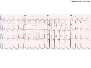
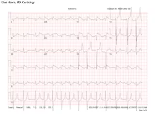
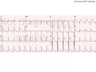
Case No. 44 • 56 years old male patient. • He is smoker, hypertensive with BMI of 27.4 • C/O of recurrent attacks of chest pain ( almost Typical ). • Clinical evaluation and resting ECG were not conclusive. • Conventional echo. study was normal • Positive myocardial perfusion study.
Comparison as regard SLSr and GLSr parameters Strain rate
Correlation between the number of affected segments in MPI and the result of CA among patient group.
Correlation between the affected segments in MPI and Strain parameters of the corresponding segments
Correlation between the affected segments in MPI and Strain rate parameters of the corresponding segments
Correlation between te number of affected segments in MPI and 2DSE parameters
Predictors of affected vessels in C.A. in relation to the strain parameters .
Predictors of affected vessels in C.A. in relation to the strain rate parameters .
Conclusions • In patients with suspected SAP, GLS assessed by 2DSE at rest can predict significant CAD. • Segmental longitudinal peak systolic strain parameters are more impaired in ischemic segments supplied by the stenotic artery. This may help in identifying which coronary artery is affected, 2DSE also is capable of identifying high-risk patients.
Conclusions • Myocardial deformation imaging based on 2-dimensional longitudinal peak systolic strain analysis may differentiate between ischemic segments and non ischemic segments compared with SPECT MPI. • It is found - and for the first time - that 2DSE is not inferior to the MPI in the non invasive diagnosis of CAD.
Recommendations • Patients with chest pain and inconclusive ECG findings can undergo echocardiography examinations with strain analysis as early as possible to facilitate early risk stratification as exclusion of significant coronary disease leads to early discharge and overall cost savings. • We recommended this study to be extended in a large scale to validate our observations.