Download

1 / 17
200 likes | 670 Views
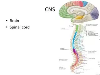
The Spinal Cord & Spinal Nerves. Together with brain forms the CNS Functions spinal cord reflexes integration (summation of inhibitory and excitatory) nerve impulses highway for upward and downward travel of sensory and motor information. Spinal Cord Protection.

E N D
The Spinal Cord & Spinal Nerves • Together with brain forms the CNS • Functions • spinal cord reflexes • integration (summation of inhibitory and excitatory) nerve impulses • highway for upward and downward travel of sensory and motor information
Spinal Cord Protection By the vertebral column, meninges, cerebrospinal fluid, and vertebral ligaments.
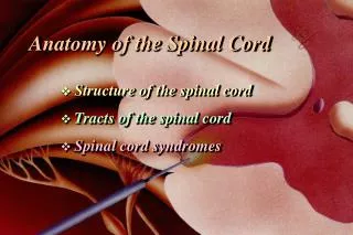
Structures Covering the Spinal Cord • Vertebrae • Epidural space filled with fat • Dura mater • dense irregular CT tube • Subdural space filled with interstitial fluid • Arachnoid = spider web of collagen fibers • Subarachnoid space = CSF • Pia mater
External Anatomy of Spinal Cord • Flattened cylinder • 16-18 Inches long & 3/4 inch diameter • In adult ends at L2 • In newborn ends at L4 • Growth of cord stops at age 5 • Cervical enlargement • upper limbs • Lumbar enlargement • lower limbs
Inferior End of Spinal Cord • Conus medullaris • cone-shaped end of spinal cord • Caudae equinae (horse’s tail) • dorsal & ventral roots of lowest spinal nerves
Spinal Cord & Spinal Nerves • Spinal nerves begin as roots • Dorsal or posterior root is incoming sensory fibers • dorsal root ganglion (swelling) = cell bodies of sensory nerves • Ventral or anterior root is outgoing motor fibers
Spinal tap or Lumbar Puncture • Technique • long needle into subarachnoid space • safe from L3 to L5 • Purpose • sampling CSF for diagnosis • injection of antibiotics, anesthetics or chemotherapy • measurement of CSF pressure
Spinal Reflexes • Automatic response to change in environment • Integration center for spinal reflexes is gray matter of spinal cord • Examples • somatic reflexes result in skeletal muscle contraction • autonomic (visceral) reflexes involve smooth & cardiac muscle and glands. • heart rate, respiration, digestion, urination, etc • Note: cranial reflexes involve cranial nerves
Reflex Arc • Specific nerve impulse pathway • 5 components of reflex arc • receptor • sensory neuron • integrating center • motor neuron • effector
Stretch Reflex (patellar reflex) • Monosynaptic,ipsilateral reflex arc • Prevents injury from over stretching because muscle contracts when it is stretched • Events of stretch reflex • muscle spindle signals stretch of muscle • motor neuron activated & muscle contracts • Brain sets muscle spindle sensitivity as it sets muscle tone (degree of muscle contraction at rest) • Reciprocal innervation (polysynaptic- interneuron) • antagonistic muscles relax as part of reflex
Flexor (withdrawal) Reflex • Step on tack (pain fibers send signal to spinal cord • Interneurons branch to different spinal cord segments • Motor fibers in several segments are activated • More than one muscle group activated to lift foot off of tack
Clinical Considerations • Checking a patient’s reflexes may help to detect disorders/injury • Plantar flexion reflex -- stroke the lateral margin of the sole • normal response is curling under the toes • abnormal response or response of children under 18 months is called Babinski sign (upward fanning of toes due to incomplete myelination in child)
Spinal Nerves • 31 Pairs of spinal nerves • Named & numbered by the cord level of their origin • 8 pairs of cervical nerves (C1 to C8) • 12 pairs of thoracic nerves (T1 to T12) • 5 pairs of lumbar nerves (L1 to L5) • 5 pairs of sacral nerves (S1 to S5) • 1 pair of coccygeal nerves • Mixed sensory & motor nerves
Dermatomes & Myotomes • Each spinal nerve contains both sensory & motor nerve fibers • Dermatome • area of skin supplied by one spinal nerve • overlap prevents loss of sensation if one damaged • sensory anesthesia requires 3 spinal nerves to be blocked • Skin on face supplied by Cranial Nerve V
Dermatomes • Damaged regions of the spinal cord can be distinguished by patterns of numbness over a dermatome region • Spinal cord transection • injury that severs the cord loss of sensation & motor control below the injury
Disorders • Neuritis • inflammation of nerves • caused by injury, vitamin deficiency or poison • Shingles • infection of peripheral nerve by chicken pox virus • causes pain, skin discoloration, line of skin blisters • Poliomyelitis • viral infection causing motor neuron death and possible death from cardiac failure or respiratory arrest