Download
1 / 15
160 likes | 307 Views
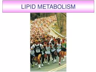
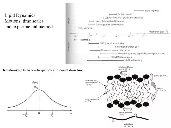
Lipid Dynamics: Motions, time scales and experimental methods. Relationship between frequency and correlation time. Models of Motion for lipids and proteins in bilayers. Conical rotation. Isotropic rotation. Isotropic rotation. Rotation along long axis. Diffusion in the plane.

E N D
Lipid Dynamics: Motions, time scales and experimental methods Relationship between frequency and correlation time
Models of Motion for lipids and proteins in bilayers Conical rotation Isotropic rotation Isotropic rotation Rotation along long axis Diffusion in the plane
Probes to measure membrane “fluidity/dynamics” Electron Paramagnetic Resonance (EPR) for monitoring membrane motions TEMPO
MI 1 ms=1/2 0 -1 N Energy O -1 0 1 ms= -1/2 EPR spectrum energy EPR spectra and hyperfine interactions
Spin labeled lipid TEMPO Isotropic tumbling Restricted motion S = order parameter Both the g-tensor and A-tensors are anisotropic
Hyperfine Tensor Directions/Definitions Director Axis and Definitions of Aperp and Apar Director axis
Examples of EPR line shapes for spin labeled lipids Mobility gradient into the bilayer
Quadrupole Lineshape for 2H, I=1 nucleus 90o 0o 2 2 2 E Quadrupole zeeman niso Need to think about 0o and 90o orientations Other faster motions average the splitting:
Oriented parallel to field Oriented perpendicular to field MLVs
NMR EPR Order parameters determined from both 2H and EPR methods From NMR relaxation studies: Keep in mind comparison between EPR and NMR: typically the 2H is less perturbing than the EPR spin probe or a fluorescent probe. But, NMR sensitivity is less. There is always a compromise.
Membrane “Fluidity” Measured by TEMPO partitioning Shows Gel to liquid transition f = fraction in the bilayer Hyperfine splitting is sensitive to polarity H is “bilayer” P is aqueous buffer