Download
1 / 2
20 likes | 39 Views
An Introduction for Quantum Dot Labeling

E N D
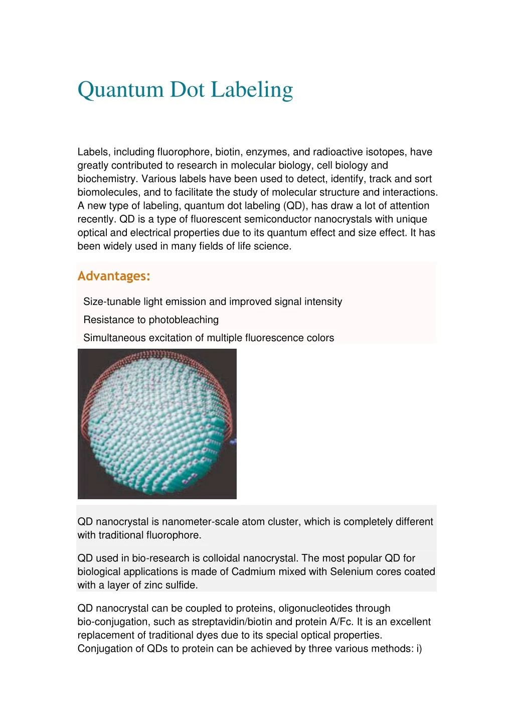
Quantum Dot Labeling INQUIRY Labels, including fluorophore, biotin, enzymes, and radioactive isotopes, have greatly contributed to research in molecular biology, cell biology and biochemistry. Various labels have been used to detect, identify, track and sort biomolecules, and to facilitate the study of molecular structure and interactions. A new type of labeling, quantum dot labeling (QD), has draw a lot of attention recently. QD is a type of fluorescent semiconductor nanocrystals with unique optical and electrical properties due to its quantum effect and size effect. It has been widely used in many fields of life science. Advantages: Size-tunable light emission and improved signal intensity Resistance to photobleaching Simultaneous excitation of multiple fluorescence colors QD nanocrystal is nanometer-scale atom cluster, which is completely different with traditional fluorophore. QD used in bio-research is colloidal nanocrystal. The most popular QD for biological applications is made of Cadmium mixed with Selenium cores coated with a layer of zinc sulfide. QD nanocrystal can be coupled to proteins, oligonucleotides through bio-conjugation, such as streptavidin/biotin and protein A/Fc. It is an excellent replacement of traditional dyes due to its special optical properties. Conjugation of QDs to protein can be achieved by three various methods: i)
use of EDC (1-ethyl-3-(3-dimethylaminopropyl) carbodiimide,); ii) direct binding using thiolated peptides or polyhistidine residues; iii) absorption or non-covalent self-assembly using engineered proteins. The emission of QD nanocrystal is narrow and symmetric, therefore allowing multiple colors to be used simultaneously, leading to its wide application in cellular labeling, tissue imaging and other biochemical assays. Profacgen provides a wide range of QD conjugation strategies to meet the specific requirements of your projects. Please feel free to contact us for more information. Reference 1 Zhao M., Zeng E., (2015). Application of functional quantum dot nanoparticles as fluorescence probes in cell labeling and tumor diagnostic imaging. Nanoscale Research Letters. 10: 171. 2 Medintz I., Uyeda H., Glodman E and Mattoussi H., (2015). Quantum dot bioconjugates for imaging, labeling and sensing. Nature Materials. 4: 435-446 INQUIRY