Download
1 / 3
30 likes | 274 Views
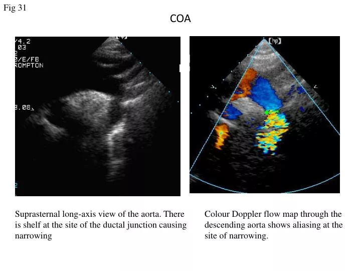
Fig 31. COA. Suprasternal long-axis view of the aorta. There is shelf at the site of the ductal junction causing narrowing. Colour Doppler flow map through the descending aorta shows aliasing at the site of narrowing. Fig 32. Spectral Doppler recording through

E N D
Fig 31 COA Suprasternal long-axis view of the aorta. There is shelf at the site of the ductal junction causing narrowing Colour Doppler flow map through the descending aorta shows aliasing at the site of narrowing.
Fig 32 Spectral Doppler recording through the descending aorta demonstrating high velocity during systole, and continued flow during diastole. Continuous flow detected in abdominal aorta.
Fig 33 Re-coarctation Continuous wave Doppler trace across the coarctation area showing a long diastolic tail. Supra-sternal view of the aortic arch in a patient with previous repaired coarctation of the aorta. Note the narrowing at the previous repaired area. The bright echo from the aortic wall suggests previous surgery.