Download
1 / 54
550 likes | 749 Views
Reactions with target molecules Cellular deregulation Repair mechanisms. “Essentials of Toxicology” by Klaassen Curtis D. and Watkins John B Chapter 3. Stages of toxicity …See Figure 3.1. 1. Delivery 2a. Interaction with target molecule 2b. Alteration of biological environment

E N D
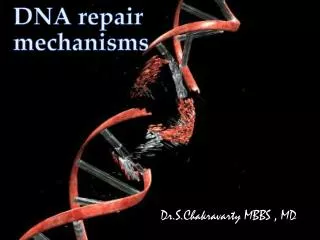
Reactions with target moleculesCellular deregulationRepair mechanisms “Essentials of Toxicology” by Klaassen Curtis D. and Watkins John B Chapter 3
Stages of toxicity…See Figure 3.1 1. Delivery 2a. Interaction with target molecule 2b. Alteration of biological environment 3. Cellular dysfunction 4. Repair or repairfailure Successful repair No Toxicity No/inadequate repair Toxicity
1. Delivery Delivery to target site Concentration at target site Elimination Distribution away from target Excretion De-activation Absorption Distribution toward target Re-absorption Activation No Toxicity Toxicity
Stages of toxicity…See Figure 3.1 1. Delivery 2a. Interaction with target molecule 2b. Alteration of biological environment 3. Cellular dysfunction 4. Repair or repairfailure
Mechanisms of toxicity Molecular targets are usually proteins, lipids, coenzymes, or nucleic acids, but rarely carbohydrates Three basic mechanisms • Formation of a stable non-covalent complex with receptor, enzyme, cofactor • Induction of a physicochemical change, e.g. pH, pO2, solvation, physical damage • Formation of reactive intermediate that binds covalently to macromolecules and/or triggers immune response
Mechanism of action Effect on specific biochemical process that leads to disruption/alteration of cellular function that eventually results in impaired physiological function (health effect) (Transient or permanent…) Symptom is the observed manifestation of a health effect (outward, macroscopic)
Mechanisms of action • Disruption or destruction of cell membrane (oxidative species, e.g. radical species) • Direct binding to cell molecule (CO+Hb; adducts, lead) • Enzyme inhibition • Cofactor • Inactivation (sequestration of cofactor) • Competition (replacement) • Binding to active site • Directly (classic enzyme inhibitors) • Indirectly: toxic metabolite binds See also Chapter 3 of Casarett and Doull’s “Toxicology”
Mechanisms of action • Secondary action: release of endogenous substance that causes damage (histamine, neuropeptides, metals displacement) • Free-radical cascade reactions (damage to proteins, DNA, lipids, mitochondria) • Structural analogue properties • Neuroendocrine context • Receptor involvement • Agonists (mimic action of endogenous substance) • Antagonists (block action of endogenous substance)
Cytochrome oxidase inhibition by cyanide stops mitochondrial respiration
Mechanism of action - dioxin http://www.stanford.edu/group/whitlock/research.html
Metabolism of bromobenzene to reactive epoxide intermediates which deplete glutathione and cause liver toxicity
Metabolism of halothane leads to direct and indirect (immune) toxicity
Redox cycling of herbicide Paraquat produces reactive oxygen species
O2 2H+ O2- . SOD HOOH O2- . O2 GSSG 2GSH 2H2O HOOH (H2O2) Coupling reactions: CAT HOOH (H2O2) 2H2O HOOH GPX
Effects of oxidative species on proteins: Oxidation of: Aminoacids targets: • cystein • methionine • tryptophan • tyrosine • sulphydryls • amines • alcohols • aldehydes Inactivation/inhibition of enzymes in cellular compartments
H2O Effects of oxidative species on lipids: • Polyunsaturated fatty acids (PUFA): • primary target of O3 peroxidation of membrane lipids • Most important mechanism of O3-induced injury • O3 + PUFA carbonyl oxide aldehydes Hydroxyhydroperoxy compound HO. H2O2 Lipid peroxidation cascade Malondialdehyde (MDA) 8-isoprostane LTB4 (PMN chemotractant) Lipid fragmentation
Effects on nucleic acids Electrophiles react with strong nucleophilic atoms of nucleic acids DNA + HO. Imidazole ring-opened purines or ring-contracted pyrimidines Strand breaks Blocked DNA replication Formation of adducts depurination (apurinic sites: mutagenic)
Reactions with target molecules • Non-covalent • Receptors • Ion channels • Enzymes • Co-factor depletion • Covalent binding • DNA • Proteins • H removal (neutral radicals) • Amino acid CH2 • Proteins • e- transfer • Hemoglobin Fe2+ hemoglobin Fe3+ (methemoglobin) • Enzymatic reactions • Protein toxins (diphtheria, cholera)
Effects on target molecules • Dysfunction • Mimics endogenous molecule • Inhibition, blocking (receptors, ion channels) • Conformational change • DNA mis-pairing • Destruction • Cross linking • Fragmentation • Oxidation/degradation (lipids)
Effects on target molecules • Antigenicity Immune response Unchanged • Dinitrobenzene • Nickel • Penicillin Following change • Quinones • Biotransformation products
Stages of toxicity…See Figure 3.1 1. Delivery 2a. Interaction with target molecule 2b. Alteration of biological environment 3. Cellular dysfunction 4. Repair or repairfailure
Alteration of biological environment • Alter pH (methanol, ethylene glycol, 2,4-dinitrophenol) • Solvents and detergents • Direct chemical effect (phosgene, sulfuric acid) • Physical space occupation (silica, asbestos, ethylene glycol, CO2)
Stages of toxicity…See Figure 3.1 1. Delivery 2a. Interaction with target molecule 2b. Alteration of biological environment 3. Cellular dysfunction 4. Repair or repairfailure
3. Cellular impairment • Cell regulation (fig. 3.6) • Gene expression • Transcription • Signal transduction (fig. 3.7) • Extracellular signal (hormone) • Cellular activity (table 3.1) • Excitable cells - neurotransmission • Other cells (Kupffer, exocrine, pancreatic)
Cellular impairment • Internal maintenance • ATP depletion (Fig. 3.8, table 3.2) Oxidative phosphorylation • Intracellular Ca+ increase (Table 3.3) Influx to cytosol • from outside (channels, membrane) • from mitochondria/ER Efflux out of cytosol • Ca+ transporters • ATPase inhibition • ROS, RNS, radicals ATP
Effects of increased cytosolic Ca+ • Inhibition of ATPase • Mito loading with Ca2+ • Dissipation of membrane potential • Reduced ATP synthesis, oxidative phosphorylation and Ca2+ cycling • Microfilament dissociation • Membrane rupture • Hydrolysis - enzyme increase • Protein, phospholipids, DNA • ROS, RNS production • Ca2+ activates dehydrogenases in citric acid cycle --> e- transport increase --> ROS, RNS
Inter-relationships Ca2+ channels that control cytosolic Ca2+ need ATP ATP Ca2+ in cytosol Mito potential Ca2+ ROS, RNS Inactivated pump
Inter-relationships Enzyme inhibition ROS, RNS ATP ONOO- DNA damage PARP NAD+
Mito Permeability Transition Ca2+ uptake Membrane potential ROS, RNS ATP MPT
Mitochondrial damage leads to cell death Pores open (1500 Da) Influx of protons, negative potential Ca2+ from mito to cytosol ATP synthesis Osmotic H20 influx Glycolysis Energy Mito swelling ATP hydrolysis Burst
Two options for cell death Robertson JD & Orrenius S. Critical Rev. Toxicology 2000, Sep; 30(5):609-27 “Molecular mechanisms of apoptosis induced by cytotoxic chemicals” http://www.roche-applied-science.com/prodinfo_fst.htm?/apoptosis
Necrosis Extensive damage All mito Multiple metabolic defects Random sequence ATP severely depleted Cell swelling and lysis Apoptosis Less extensive Some/many mito Some metabolic defects Ordered sequence Some ATP available Cell shrinkage, membrane bound fragments MTP - cell death
Stages of toxicity…See Figure 3.1 1. Delivery 2a. Interaction with target molecule 2b. Alteration of biological environment 3. Cellular dysfunction 4. Repair or repair failure
Molecular repair • Proteins reduction (re-activation) NADPH • Protein refolding (heat-shock proteins) • Protein degradation and re-synthesis • Lipid reduction (GPO, GR, NADPH) • DNA repair
DNA damage repair • Direct: photolyase (UV-dimers, O6-methyl-G removal) • Excision DNA glycosylase (removes AP site) AP endonuclease (PO3 bond) DNA polymerase (replicates sequence) Ligase (ties the ends) PARP (multiple ADP ribose - unfolds/facilitates repair) • Recombination Sister chromatid exchange
Cellular/Tissue repair • Single cell - regeneration (neurons) • Tissue • Apoptosis • Proliferation • Chemokine priming (G0-G1) :TNFa, IL-6 • Chemokine progression (G1-GM) :HGF, TGFa • Migration • ECM (Stellate cells, PDGF, TGFb)
Inflammation Macro’s IL-1, TNFa endothelia, fibroblasts Vascular dilation Leukocyte infiltration Release of PAF, LTB4, cytokines Leuko-endo adhesion
L-citruline H+ NO2. NOS L-arginine + O2 NO. Fenton HO. Oxidase NAD(P)H + O2 O2. NAD(P)+ H+ H20 O2 Cl- MPO HOOH + H+ +Cl- HOCl Side reactions - Inflammatory oxidative burst • Three pathways of HO. generation: • NAD(P)H oxidase (macro’s and granulo’s) • Nitric oxide synthase (NOS) (macro’s) • Myeloperoxidase (MPO) (granulo’s)
More side reactions • Gene expression • Cytokines IL-6, IL-1, TNFa • Acute phase proteins • Minimize injury • Facilitate repair (inhibit lysosomal proteases) • Plasma proteins • CYP450, GSTs (detox) • Generalized reactions • Fever (IL-1, IL-6, TNFa) hypothalamus • Pituitary (ACTH --> cortisol) (negative feedback)