Download
1 / 60
610 likes | 648 Views
Learn about the maturation process, classification, shapes, and abnormalities of erythrocytes (red blood cells) in the laboratory. Understand the significance of cell arrangement, color, size, and shape in RBC analysis.

E N D
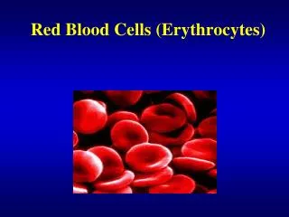
Abnormalities in Erythrocytes Laboratory Procedures
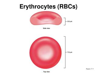
Red Blood Cells:aka ___________ • ______________is the process of maturation of a RBC • Formed by the stem cell through action of the _________________________called ___________(EPO) • Maturation of a RBC takes about 5 days.
Erythrocyte Life Span Stem Cell → _________________→ Prorubricyte → ___________________→ Metarubricyte→ ________________________→ RBC Metarubricyte- nucleated RBC released in severe anemia.
Erythrocyte Life Spans • Dog ~________ days • Cat ~_______ days • Cow ~ 160 days • Horse ~ 145 days • Man ~ 120 days • Mouse ~ 30 days
RBC’s continued • No _______________ • ____________ varies among species
Classification of RBC’s • RBC’s are classified the following criteria: • 1) Cell _________________ on blood film • 2) __________ • 3) __________ • 4) __________ • 5) Presence of ___________ on erythrocytes (We will come back to this one!)
Classification of RBC’s1) Cell Arrangement on Blood Film • Normal erythrocytes should lie in a nice, even, __________ layer on the _____________-most edge of a blood film
Rouleaux Formation • Defined:________________________________________________. • It can be a sign of increased fibrinogen or globulin concentration • Can be an _______seen in blood that is held too long before preparation of blood slide or if refrigerated. • If a drop of _______ is added to blood, rouleaux will disperse
AgglutinationOr Auto-agglutination • May appear as rouleaux (stacks) or in clusters • Occurs in ______________disorders • An ______________ coats the cell causing bridging or clumping. • If a drop of _____________ is added to blood, agglutination will not disperse
Classification of Blood Cells • 2) Color of Erythrocyte • Erythrocytes that are normally colored are called _______________________________ • Polychromasia: ______________________________ • Polychromasia can appear as ______chromasia or _____chromasia.
Polychromasia • Can exist as ________________________ or ________________________. • Polychromatic erythrocytes exhibit a ____________ tint. This is caused by a small amount of ______________ retained in the cytoplasm. These may appear as a ________________________________________. (We will talk about these later in this presentation.)
Hypochromasia • Is a __________________ in color, due to a decreased staining intensity caused by insufficient ________________________ within the cell. • Most commonly caused by __________________________________
Hyperchromasia • Refers to a cell that appears _____________ than normal cells. This gives the appearance that the cell is over-saturated with _______________________. • TRUE hyperchromasia cannot exist! • Each erythrocyte has a ____________________________________________ for hemoglobin and over-saturation cannot occur • If cells appear hyperchromic, there is another underlying concern.
MCHC • Stands for: ______________________________________________ • Describes cells as normochromic or hypochromic (why no hyperchromic?) • Normal MCHC is 31-36% • (You will learn this calculation in Clin. Path)
Classification of RBC: 3) Size of Erythrocyte • Erythrocytes that are of normal, consistent size are called ___________________________________ • Anisocytosis: _______________________________ • ___________________ cells are smaller than normal cells • ___________________ cells are larger than normal cells
Anisocytosis • Variations in ___________________ • Can indicate ___________________ • Classified by ____________________ OR _____________________
Macrocytosis • Simply means that there are an abnormal amount of cells _____________________ than normal size. • Can indicate __________________ disease or _____________________ deficiency.
Microcytosis • Indicates that there is an abnormal amount of cells that are ___________________ than normal. • Can indicate _______________ deficiency.
MCV • Stands for ____________________________________________ • Describes cells as being ____________________, ___________________, or _____________________. • Measures the average volume of RBC’s. • Normal values are 66-77 fL (femtolitres)
Classification of RBC’s4) Shape of Erythrocyte • Poikilocytosis: ______________________________ • This is an “________________________” term that cannot suggest a ______________________
Poikilocytosis • Is a major deviation in the normal ____________ of the erythrocyte. • Remember that this term is an umbrella term for abnormally ________________ erythrocytes, and should not be used as a _______________.
Many Poikilocytes • All of the following cells are under the “umbrella” of Poikilocytes. They just have different names! • (Remember rule #2)
Schistocytes (Fragmented Cells) RBC’s with abnormal shape. Formed as a result of shearing of the cell by _______strands. This occurs when red blood cells rapidly pass through microvasculature that is lined or meshed with strands. They are observed in fragmentation hemolysis caused by _______________, ________________, ______________, and possibly ________________________anemia.
Acanthocytes (Spur Cells) • The term acanthocyte is derived from the Greek word “acanthi” meaning “thorn” Acanthocytes are cells with five to ten irregular, blunt, finger-like projections. • The projections with vary in ______, __________ ,and surface ____________________. • These cells are seen in animals with altered ________________metabolism such as cats with _______________________________ or dogs with ______________ disease.
Echinocytes(Burr Cell) • Echinocytes have multiple, small, delicate regular shaped spines _______________________________ around the cell and are indistinguishable from artificially ___________________cells.
EchinocytesContinued • Echinocyte formation can be ___________________, often seen with slow drying blood films or if the EDTA tube was ______________. This artifact is then termed ____________________. • Echinocytes have been associated with _______________disease, lymphosarcoma and rattlesnake bites in dogs. • They can been seen after exercise in ________________.
Crenation Identified as the presence of many __________ membrane projections involving most RBC’s. It is usually an ___________due to slow drying of the blood film. Commonly observed in ___________blood but can be seen in any species.
Drepanocytes (Sickle cell) • These cells are _________________ shaped with pointed ends. • Drepanocytes are often seen in normal blood of __________and ________________. • It is thought to be a result of low _______________tension.
Pre-keratocytes • Cells with ____________________are called _______________cells or pre-keratocytes.
Keratocyte (Helmet Cells) • Also called ___________cells. Keratocytes are associated with trauma especially cellular damage from contact with ________________strands.
Spherocytes Cells have a spheroid shape instead of the usual biconcave disk shape. Have ______________cell membrane and are ___________________________. Seen most frequently in_______________ _________________________________(AIHA). Usually seen in _______.
Dacryocytes (tear drop cells) • These tear drop shaped cells are seen in _____________________________diseases. • These cells, when produced as an ______________can be identified by the direction of their tail.
Dacryocytes produced as an artifact have their tails pointing in ___________________________________________.
Codocytes (aka Leptocytes) • Is an “___________ term” describing cells that are characterized by an increase in membrane ___________________________. • Include the following: • ________________________________ • ________________________________
Folded Cells and Stomatocytes • The appearance of Folded cells with their _____________________________central pallor has been compared to a fish mouth and a coin slot. • Stomatocytes resemble a ____________________ • Both are associated with a hereditary condition but are also seen in ____________disease, acute alcoholism (humans), and ________________imbalances.
Folded Cells Stomatocytes
Target Cells Observed mainly in _______________. Represent cells with an ____________________ membrane-to-volume ratio not specific to any disease. The ________________________________is thin and flimsy.
Target Cells (Bull’s Eye Cells) • Thin, bell-shaped cells • Centrally stained area • Can indicate ____________disease or hemoglobinopathies. • May be seen as ________________when smears made in high ______________ or if ______________ dry.


![ERYTHROCYTES [RBCs]](https://cdn3.slideserve.com/5385917/erythrocytes-rbcs-dt.jpg)
![ERYTHROCYTES [RBCs]](https://cdn3.slideserve.com/6667616/erythrocytes-rbcs-dt.jpg)