Download
1 / 78
880 likes | 1.35k Views
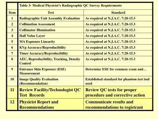
The ACR CT Accreditation Program and the Medical Physicist. Maynard High, PhD New York Medical College with the most kind assistance of Cynthia McCollough, PhD Mayo Clinic. Aim of CT Accreditation. Peer Review and Evaluation of facility, including: personnel qualifications image quality

E N D
The ACR CT Accreditation Program and the Medical Physicist Maynard High, PhD New York Medical College with the most kind assistance of Cynthia McCollough, PhD Mayo Clinic
Aim of CT Accreditation • Peer Review and Evaluation of facility, including: • personnel qualifications • image quality • quality control procedures • patient radiation exposure
Positive Outcomes of CT Accreditation Process from the Viewpoint of a Satisfied User • Technologist: • involvement in performance evaluation • Technologist, radiologist, and physicist: • increased awareness of patient dose • Patient: • review and possible modification of scan protocols affecting dose and image quality
Financials of CT Accreditation • Costs are not insignificant: • ~ $5K for 1 CT site with phantom • Some insurers now require CT accreditation: • Many more insurers will require CT accreditation within a year:
CT PROGRAM STATISTICS April 2003 • 200 applications received • 40 facilities accredited • ??% passed on 1st attempt • too early in program for statistics • failures about 50% clinical / 50% phantom
The Application Process • Submission of site info and personnel credentials • Acquisition of ACR CT phantom • Submission of CT data and images • clinical images • phantom images • dose measurements
Personnel Credentials • Radiologist: • Competence, continuing experience, CME • Technologist: • Competence, CME • Medical Physicist: • Competence, CME
Medical Physicist Credentials 1)RECOMMEND ABR certification in • Diagnostic Radiological Physics or • Radiological Physics 2)Be Familiar with CT 3)Be in accordance with ACR Standard for CME • no specific requirement for CT CME
ACR Standard for CME:Medical Physicist • 150 hrs/3 yrs (Category 1,2 and MEP) • this 150 hrs includes the 60 hrs below • Category 2 includes meeting attendance, self-study, teaching, publications,peer-review activities, etc • 60 hrs/3 years (Category 1 or MEP)
The Application Process • Submission of site info and personnel credentials • Acquisition of ACR CT phantom • Submission of CT data and images • clinical images • phantom images • dose measurements
ACR CT Phantom Information Courtesy of CYNTHIA McCOLLOUGH, PHD • All slides of this format and color scheme were kindly loaned to me for use in this talk by Cynthia McCollough, PhD, Chairperson of the ACR CT Accreditation Physics Subcommittee
Physics Subcommittee • Cynthia McCollough, Ph.D., Chair • Tom Payne, Ph.D. • Mike McNitt-Gray, Ph.D. • Tom Ruckdeschel, M.S. • Jim Brink, M.D. • ACR: Pam Wilcox, Penny Butler, Krista Bush, Chris Riha
Consensus Opinion re: Accreditation Phantom and Film • Single phantom design must be used • No existing phantom had all desirable test objects • Solid one-piece construction, 20-cm diameter • Test objects must be simple to evaluate • Objects and tests must be extendable to spiral • Dosimetry will be CTDI100-based in PMMA • Assessment of phantom images will be film-based until ACR converts to digital submission process
The ACR CT phantom was designed to examine a broad range of scanner parameters • Positioning Accuracy • CT # Accuracy • Slice Width Accuracy • Low Contrast Resolution • High Contrast (Spatial) Resolution • Image Uniformity and Noise • Image Artifacts • Distance Measurement Accuracy • Section Sensitivity Profiles
Align light accuracy Align table to gantry Table/gantry tilt Scout slice localization accuracy Table incr. accuracy Slice thickness CT# accuracy/linearity Hard-copy display Image Quality high contrast resolution low contrast resolution image uniformity noise artifact evaluation CTDI Patient dose for exams ACR Accreditation Application Tests
Align light accuracy Align table to gantry Table/gantry tilt Scout slice localization accuracy Table incr. accuracy Slice thickness CT# accuracy/linearity Video display Hard-copy display Image Quality high contrast resolution low contrast resolution image uniformity noise artifact evaluation CTDI Patient dose for exams Safety evaluation Required State tests Physicist’s Annual CT Survey (ACR)
Align light accuracy Slice thickness CT# accuracy Video display Hard-copy display Image Quality high contrast resolution low contrast resolution image uniformity noise artifact evaluation Technologist CT QC (ACR)
Head Foot 4 3 2 1 High contrast resolution Uniformity & noise Distance accuracy & SSP Low contrast resolution Alignment CT # Slice width 20 cm 4 cm
Technologist and Phantom Testing • Try to give technologist responsibility for accreditation phantom testing • best done together with physicist • helps technologist better understand the physicist’s annual survey (ACR required) • helps the physicist understand how technologist sets up protocols (useful information for annual survey)
Site Scanning Data Form • Site or manufacturer-recommended protocols • Adult Head: Routine head CT for evaluation of patient with headaches to exclude neoplasms • High Resolution Chest: CT exam of the chest for evaluation of diffuse lung disease • Adult Abdomen: Routine oncologic CT survey of the abdomen for detection of possible liver metastases • Pediatric Abdomen: CT examination of pediatric (approx. 5 years old) abdomen for the evaluation of blunt trauma injuries
Instruction Manual • Detailed step by step scan and analysis instructions • Tells which set of scan parameters to use to acquire which phantom images • Data sheet provides cells for measured and calculated data • W/L and location on film grid given for all images to be filmed • Dose measurement and calculation methods
Test 1: Phantom and Scanner Alignment • Align Module 1 to lasers • Scan with Hi Res Chest protocol • Prescribe a scan at center of Module 4 ie, 120 mm superior • Scan with Hi Res Chest protocol
MODULE 1 Polyethylene ≈ -97 HU “Bone” ≈ +910 HU Water ≈ 0 HU Acrylic ≈ +120 HU Air ≈ -1000 HU
High-resolution chest technique Must see all four BBs (in Modules 1 & 4) Longer wire must have same number of lines above and below (±1) Wires are 0.5 mm apart in z-direction WW = 1000 WL = 0
Test 2: CT Number Calibration and Slice Thickness • Align Module 1 to lasers • Scan with Adult Adomen protocol & all kVp’s available • Record all HU and slice thickness • Also scan with 3, 5, 7 mm & hi res chest slice thickness • Record water HU and slice thickness for each scan.
“Bone” Poly Water Air Acrylic WW = 400 WL = 0 Adult abdomen technique CT number measured in 5 materials CT number of water measured at several scan widths and kVps
Adult abdomen technique Scan width measured at several scan widths 11/2 = 5.5 10/2 = 5 WW = 400 WL = 0
Test 3: Low Contrast Resolution • Scan Module 2 with Abdomen and Head protocols • Record the smallest rod seen
Low contrast = 6 HU ± 0.5 HU MODULE 2 2 mm 25 mm 3 mm 6 mm 4 mm 5 mm
Adult abdomen and head techniques Record diameter of the smallest set of rods for which all 4 rods can be seen ROI check of absolute contrast using large rod WW = 100 WL = 100
Test 4: Uniformity, Noise, Artifacts, and Distance Accuracy • Scan Module 3 with Abdomen protocol • Record HU & SD in center • Record HU at 12, 3, 6, 9 o’clock • Examine for artifacts and record • Check distance accuracy (optional)
MODULE 3 100 mm
Adult abdomen and head techniques Measure uniformity and noise Assess for artifacts Measure distance accuracy (optional) WW = 100 WL = 0
Test 5: High Contrast Resolution • Scan Module 4: • Hi Resolution Chest protocol • Abdomen protocol • Record limiting lp/mm
Bar patterns: lp/cm MODULE 4 12 4 10 5 9 6 8 7
Adult abdomen, adult head and high-resolution chest techniques Record the first highest frequency bar pattern for which the bars and spaces merge WW = 100 WL 1100
CT ART? At soft-tissue window settings these streaks are visible and not of concern WW = 400 WL = 0
The Application Process • Submission of site info and personnel credentials • Acquisition of ACR CT phantom • Submission of CT data and images • clinical images • phantom images • dose measurements
Dose Information for ACR Accreditation • Submit calculations of CTDIvol, DLP and Effective Dose using the site’s measured CTDIw and the reported scan acquisition parameters (pitch) • Routine head (cerebrum) • Adult abdomen • Pediatric abdomen (5 y.o) • CDTI phantom acquisition must be filmed