Download
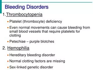
1 / 66
760 likes | 1.74k Views
Hemophilia and Rare Bleeding Disorders. 2007 EMA Advanced Emergency & Acute Care Medicine Conference Atlantic City, NJ September 24, 2007. Edward P. Sloan, MD, MPH FACEP Professor Department of Emergency Medicine University of Illinois College of Medicine Chicago, IL.

E N D
2007 EMA Advanced Emergency & Acute Care Medicine ConferenceAtlantic City, NJSeptember 24, 2007
Edward P. Sloan, MD, MPH FACEPProfessorDepartment of Emergency MedicineUniversity of Illinois College of MedicineChicago, IL
Attending PhysicianEmergency MedicineUniversity of Illinois HospitalOur Lady of the Resurrection HospitalChicago, IL
Disclosures • Novo Nordisk grant to conference • FERNE Chairman and President • FERNE grants from Novo Nordisk • No financial disclosures • eMedicine source materials • Slide materials from Novo Nordisk
Global Objectives • Maximize patient outcome • Utilize health care resources well • Optimize evidence-based medicine • Enhance ED practice
Sessions Objectives • Learn about hemophilia and RBDs • What are the diseases? • How do patients present? • What are management principles? • What specific therapies? • How to enhance pt outcomes?
Case Presentation 17 year old presents to ED Known hemophilia A Fell off of bicycle Abdominal trauma Hypotensive, tachycardic Abdominal tenderness What do you do?
ED Bleeding Disorder Patients: Key Concepts Identify the bleeding disorder Establish if bleeding is present Treat the bleeding Treat the bleeding disorder Establish endpoint for Rx success Disposition based on Dx, Rx, risk
Background Rare disorder: Affects fewer than 200,000 Americans (NIH office of Rare Diseases) Hemophilia Other bleeding disorders Rare Bleeding Disorders
Hemophilia Hemophilia A Congenital deficiency of factor VIII (FVIII) Hemophilia B: Christmas Disease Congenital deficiency of factor IX (FIX)
Hemophilia Insufficient generation of thrombin by FVIIIa and FIXa complex through the intrinsic pathway of the coagulation cascade
Fibrin Clot Structure Hemophilia A Normal Clot Structure
Hemophilia Severity Based on procoagulant levels or bleeding severity Severe: <1% clotting factor present Moderately severe: 1-5% Mild: 5-40% Clinical bleeding severity may not match amount of deficiency
Other Inherited Bleeding Disorders Congenital factor deficiencies von Willebrand’s Disease Other congenital platelet disorders Glanzmann’s Thrombasthenia Bernard Soulier Syndrome
von Willebrand’s Disease Autosomally inherited bleeding disorder, mucocutaneous Deficiency or dysfunction of the protein termed von Willebrand factor (vWF) Primary hemostasis is impaired Defective interaction between platelets and the vessel wall
Factor VII Deficiency Fewer than 200 cases of true factor VII deficiency have been reported Gene mutations, protein dysfunction Factor VII coagulant activities measured in the laboratory are not well correlated with bleeding manifestations
Acquired Bleeding Disorders Vitamin K Deficiency Severe Liver Disease Factors II, VII, IX and X are decreased Platelets dysfunctional Renal Disease Platelet dysfunction
Acquired Bleeding Disorders Oral Anticoagulant Therapy Prolonged Use of Antibiotics Develop anti-platelet antibodies Vitamin K deficiency Acquired Inhibitors (Antibodies) Post malignancy Related to pregnancy Idiopathic Elderly
Patient Demographics Hemophilia Present in childhood, esp with greater disease severity All races X-linked, recessive males
Patient Presentations Mannucci et al. Blood 2004;104:1243-1252
History What is the Bleeding Disorder? vWD, Hemophilia A/B, other factor deficiency or platelet disorder
History What is the severity of the factor deficiency? Severe - < 1% factor present Bleed spontaneously and often e.g. weekly Moderate – 1-5 % Can have spontaneous bleeding but less frequent e.g. monthly Mild - > 5 % Bleed only when hemostasis is challenged e.g. trauma and surgery
History Do they have a inhibitor (assoc with congenital factor def)? What is their HIV/Hepatitis Status? How is the bleeding disorder being treated? When was your most recent treatment or infusion? Are you taking other medications?
Hemorrhage History General - Weakness and orthostasis Musculoskeletal (joints) - Tingling, cracking, warmth, pain, stiffness, and refusal to use joint (children) CNS - Headache, stiff neck, vomiting, lethargy, irritability, and spinal cord syndromes
Hemorrhage History GI - Hematemesis, melena, frank red blood per rectum, and abdominal pain Genitourinary - Hematuria, renal colic, and postcircumcision bleeding
Hemorrhage History Other - Epistaxis, oral mucosal hemorrhage, hemoptysis, dyspnea (hematoma leading to airway obstruction), compartment syndrome symptoms, and contusions; excessive bleeding with routine dental procedures
Physical Exam General hemorrhage signs Organ-specific hemorrhage signs Hepatitis signs Infections signs Medic Alert bracelet Wallet
Physical Exam General hemorrhage signs Organ-specific hemorrhage signs Hepatitis signs Infections signs Medic Alert bracelet Wallet (Hemophilia Treatment Center Card)
Laboratory Testing CBC (Hb, platelets, WBC) PT, aPTT vWF:Ag (Von Willibrand factor antigen) Ristocetin Co-Factor Measures vWF activity to identify qualitative vWF disorder Factor coagulant activity e.g. VIII:C, IX:C in hemophilias Bleeding time?
Laboratory Testing PT: Extrinsic, should be normal unless FVII deficiency or acquired aPTT: Intrinsic, elevated in moderate hemophilia disease severity
Other ED Testing Extremity xrays Head CT Abdominal CT Tests for increased compartment pressures Nuclear bleeding studies
Initial Management Treat the patient ABCs Direct hemorrhage control Hemodynamic support Crystalloids Blood products Specifically assist hemostasis
The 3 Phases of Hemostasis Primary hemostasis: • Vasoconstriction • Platelet adhesion • Platelet aggregation and contraction Secondary hemostasis: • Activation of coagulation factors • Formation of fibrin Fibrinolysis: • Activation of fibrinolysis • Lysis of the plug
Blood Vessel & Endothelium • Hemostasis requires and involves various physiological components: • The blood vessel wall • Endothelial cells • Subendothelial tissue • Smooth muscle cells • The components of blood • Platelets (thrombocytes) • Coagulation (clotting) factors • Fibrinolytic/ anticoagulant proteins
Primary Hemostasis: Vasoconstriction • The first response to endothelial injury is the constriction of the damaged vessel which reduces the blood flow at the site of injury
Primary Hemostasis: Formation of a Platelet Plug • The exposure of subendothelial components such as collagen promotes platelet adhesion • The adherence of platelets to the sub-endothelium leads to platelet activation and the formation of platelet aggregates (platelet plug)
Secondary Hemostasis • At the site of vascular injury binding of endogenous factor VII/VIIa to tissue factor (TF) leads to the generation of small amounts of thrombin • Thrombin activates platelets and additional coagulation factors which subsequently generate large amounts of thrombin • This “thrombin burst” induces the generationof a haemostatic plugthat prevents furtherblood loss Adapted from Hoffman M et al., 2001.1
How a Blood Clot Forms: Step 2 PTT PT HMWK,PK XII TF XI IX VIIa/TF VII VIIIa X X Va IIa (thrombin) II (prothrombin) fibrinogen Fibrin Clot (factor I)
What is Broken? Platelets Clotting factors Coagulation cascade
What Can Be Provided? Vitamin K FFP (Fresh frozen plasma) PCC (prothrombin complex concentrate) Platelets, packed RBCs, whole blood Specific clotting factors Anti-fibrinolytics, anti-hemophilics
Hemophilia A: Factor VIII Recombinant factor VIII concentrate is the preferred source of factor VIII. The factor VIII activity level should be corrected to 100% of normal for potentially serious hemorrhage. Units factor VIII=(weight in kg)(50 mL plasma/kg)(1 U factor VIII/mL plasma)(desired factor VIII level minus the native factor VIII level)
Hemophilia A: Factor VIII As an example, an 80-kg individual diagnosed with hemophilia with known 1% factor VIII activity level presents to the ED with a severe upper GI bleed. Units factor VIII = (80 kg)(50 mL/kg)(1 U factor VIII/mL)(.99) = 3960
Hemophilia A: Factor VIII Next dose: 12 hours later, 1/2 initial dose. Minor hemorrhage: 1-3 doses factor VIII. Major hemorrhage: many doses, continued factor VIII activity monitoring. Goal: trough activity level at least 50%.