Download
1 / 56
741 likes | 1.48k Views
Facial Trauma. Babak Saedi MD Otolaryngologist Tehran University of Medical Sciences. The External Bony Facial Skeleton. Composed mainly of the frontal bone, temporal bones, nasal bone, zygomas , maxilla, and mandible.

E N D
FacialTrauma Babak Saedi MD Otolaryngologist Tehran University of Medical Sciences
The External Bony Facial Skeleton • Composed mainly of the frontal bone, temporal bones, nasal bone, zygomas, maxilla, and mandible. • Ethmoid, lacrimal, sphenoid bones contribute to inner portion of orbits • Upper third - above superior orbital rim • Middle third (midface)- superior orbital rim down through maxillary teeth • Lower third - mandible
Maxillofacial Trauma Patient evaluation
Patient Evaluation • History • Physical exam • Other systems: - Airway - Circulation - CNS (GCS)
Physicalexamination • Orbit • Nasal airway • Dental occlusion • Neurovascular
Softtissuedamage • Contusion • Avulsion • Laceration (loss of soft tissue – penetrating trauma)
Physical Examination • First, inspect face for deformity and asymmetry • Enophthalmos, proptosis, ocular integrity, ocular movements • Nasal septum for position, integrity, and presence of septal hematoma • Epistaxis or CSF rhinorrhea
Physical Examination • Complete neurological exam must be performed on any patient with suspected facial trauma • Sensation - test all 3 major branches of the trigeminal nerve • Motor function - assess facial nerve by having patient wrinkle forehead, smile, bare teeth, and close eyes tightly
Physical Examination • Palpation of facial structures - the infraorbital and supraorbital ridges, zygoma, nasal bones, lower maxilla, and mandible • Assess for tenderness, bony deformities, crepitus, . . . • Malocclusion or step-off in dentition may be sign of mandibular fracture
Diagnostic Imaging • Should focus on bony integrity, fluid-filled sinuses, herniation of orbital contents, and subcutaneous air • Overall status of the patient, physical exam findings, and the clinician’s initial impression determine timing and nature of imaging ordered
Plain films • Traditionally the mainstay in the radiographic evaluation of facial trauma • Standard plain film facial series: Waters (occipitomental), Caldwell (occipitofrontal), and lateral views • Panoramic films are used to best evaluate mandibular fractures
CT scan • Offers a viable, cost-effective alternative to plain films • Very helpful in the evaluation of facial trauma when facial edema, lacerations, other injuries, or altered level of consciousness limit usefulness of clinical exam
MRI • Limited role of MR in evaluation of facial trauma due to insensitivity of MR to fractures • Used to provide complimentary information to CT in the evaluation of the eye and its associated structures
Nasal Fractures • Most common site of facial trauma due to location • May be displaced medialy, laterally or posteriorly • Requires control of epistaxis and drainage of septal hematoma, if present
Nasal fractures - classification • Class 1 - frontal or frontolateral trauma - vertical septal fracture - depressed or displaced distal part of nasal bones • Class 2 - lateral trauma - horizontal or C-shaped septal fracture - bony or cartilaginous septum fracture - frontal process of maxilla fracture
Nasal fractures - classification • Class 3 - high velocity trauma - fracture extends to ethmoid labyrinth - bony septum rotates posteriorly - bridge collapse - upturned tip, revealing nostrils - depressed nasal bones pushed up under frontal bones - apparent inter-ocular space widening
Nasal fracture • Diagnosis: - physical exam (asymmetry, deviation, epistaxis, swelling, . . .) • Radiography: - do not have a role in management • Timing: - before 10 days to 2 weeks - within two hours after injury
Nasal fracture • Managements: (closed & open reduction) • Complications: - septal hematoma - CSF leakage - ophthalmologic compl.
Zygomatic Fractures • Tripod fracture: zygomaticofrontal suture, zygomaticotemporal suture, and infraorbital foramen • Present with flatness of the cheek, anesthesia in the distribution of the infraorbital nerve, diplopia, or palpable step defect
Maxillary Fractures • Le Fort I – maxilla • Le Fort II – maxilla, nasal bones, and medial aspects of orbits (pyramidal disjunction) • Le Fort III – maxilla, zygoma, nasal bones, ethmoids, vomer, and all lesser bones of the cranial base (craniofacial disjunction) • Usually in combination
Blowout Fracture of the Orbit • Fractures of the orbital floor may occur with orbital wall fractures or as an isolated injury. • When the orbital floor, being the weakest area, herniation of orbital contents down into the maxillary sinus may occur (hanging drop sign). • Patients may present with enophthalmos, impaired ocular motility, diplopia due to entrapment of the inferior rectus muscle within the fracture fragments, and infraorbital hypoesthesia.
Maxillofacial Trauma-Specific Fractures • Orbital Fractures • Usually through floor or medial wall • Enophthalmos • Anesthesia • Diplopia • Infraorbital stepoff deformity • Subcutaneous emphysema
Blowout Fracture of the Orbit • This child presented with diplopia following blunt trauma to the right eye. On exam, he was unable to move his right eyeball up on upward gaze.
CT: Blowout Fracture of Orbit • A: Orbital blowout fracture with displacement of the floor (arrow), distortion of the inferior rectus, and herniation of orbital fat through defect. Arrowhead indicates medial fracture. • B: Note opacified left anterior ethmoid air cells and displaced medial orbital fracture (arrowheads).
Maxillofacial Trauma-Specific Fractures • Frontal Sinus/Bone Fractures • Direct blow • Frequent intracranial injuries • Mucopyoceles • Consult with NS for treatment, disposition and antibiotics • Nasoethmoidal-Orbital Injuries • Lacrimal apparatus disruption • Bimanual palpation if medial canthus pain • CT face
Maxillofacial Trauma-Specific Fractures • Orbital Fissure Syndrome • Fracture of the orbital canal • Extraocular motor palsies and blindness • If significant retrobulbar hemorrhage, may need cantholysis to save vision • Zygomatic Fractures • Tripod fracture • Most serious • Lateral subconjunctival hemorrhage • Need ORIF • Arch fracture • Most common • Outpatient repair
Maxillofacial Trauma-Specific Facial Fractures • Mandibular Fractures • Second most common facial fracture • Often multiple • Malocclusion • Intraoral lacerations • Sublingual ecchymosis • Nerve injury • Plain films • Panorex • CT • Open Fractures • Prophylactic Ab.
Types of fracture • Simple • Greenstick fracture (rare, exclusively in children) • Fracture with no displacement (Linear) • Fracture with minimal displacement • Displaced fracture • Comminuted fracture Extensive breakage with possible bone and soft tissue loss • Compound fracture Severe and tooth bearing area fractures • Pathological fracture (osteomyelities, neoplasm and generalized skeletal disease)
Favourable or unfavourable • They can be vertically or horizontally in direction • They are influenced by the medial pterygoid-masseter “sling” • If the vertical direction of the fracture favours the unopposed action of medial pterygoid muscle, the posterior fragment will be pulled lingually • If the horizontal direction of the fracture favours the unopposed action of messeter and pterygoid muscles in upward direction, the posterior fragment will be pulled lingually • Favourable fracture line makes the reduced fragment easier to stabilize
Panoramic X-Ray Film of the Mandible • Note fractures in left angle and right body of mandible • Multiple fractures are present more than 50% of the time and are usually on contralateral sides of the symphysis
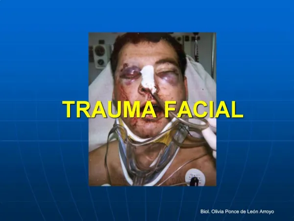
Approach to the Patient with Traumatic Injury of the Face • Facial trauma is defined as injury to the soft tissues of the face (including the ears) and to the facial bony structures. • May result in hemorrhage and airway obstruction accompanied by multisystem involvement (as many as 60% of patients have associated injuries) • Evaluation includes history, physical exam, and diagnostic imaging
Principles of treatmentsimilar to elsewhere fractures in the body • Reduction of fragments in good position • Immobilization until bony union occurs These are achieved by: • Close reduction and immobilization • Open reduction and rigid fixation Other objective of mandible fracture treatment: • Control of bleeding • Control of infection
Treatment options • No treatment • Soft diet • Maxillomandibular fixation • Open reduction - non-rigid fixation • Open reduction - rigid fixation • External pin fixation • Lag screw
Close reduction • Arch bars ▶IMF prior to rigid fixation ▶For the purpose of close reduction