Download
1 / 32
320 likes | 548 Views
Fitting Fibrils A geometrical approach to plant cell wall development. Introduction to plant cell wall morphology The current paradigm The geometrical theory Results Conclusions. Bela Mulder FOM Institute AMOLF Amsterdam, NL.

E N D
Fitting FibrilsA geometrical approach to plant cell wall development • Introduction to plant cell wall morphology • The current paradigm • The geometrical theory • Results • Conclusions Bela MulderFOM Institute AMOLFAmsterdam, NL Anne-Mie EmonsMiriam AkkermanPlant Cell BiologyWageningen University, NL
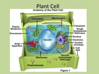
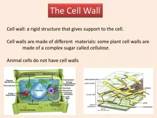
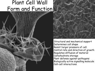
Why study the cell wall ? Cell walls provide protection and allow plants to exploit turgor pressure to raise themselves against gravity • Plants make up 99% of the biomass of earth. • 10% of this biomass is fixed in plant cells. • Important source of raw materials: wood, paper, fibers …
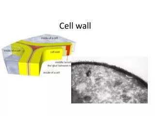
Zooming in on the cell wall Example cell: root hair Cross-section
Shadow-cast EM image Surface section
Cellulose microfibrils in a polysaccharide matrix 2-4 nm CMF
Real world analogues Fibre-laminates are both tough and flexible Helicoidally wound strings pack efficiently
The mechanism of CMF synthesis Certain: CMF synthases channel UDP glucose from inside the cell, and “spin” the CMF. Brown et al. (1997)Arioli et al. (1999) Plausible: The CMF synthases are propelled forward by the polymerization force and move in the plasma membrane.
The current paradigm The so-called microtubule/microfibril paradigm (Giddins&Staehelin [1991]) The CMF synthases are “guided” by the cortical microtubules.
However … • The hypothesis is mainly supported by the the co-alignment of CMFs and MTs in expanding cells, where forces are exerted. • In many non-expanding cells there is no co-alignment between MTs and CMFs (Emons [1983,->]) • New Arabidopsis mutants show normal wall development even when the cortical MT organization is disrupted (Wasteneys et al.) • It begs the question of how the cortical MTs are (re)organized.
Background to the geometrical model CMFs are deposited by CMF synthases that move in the plasma membrane. CMF cell interior Deposition takes place in the limited space between the cell membrane and the already extant wall. existing wall synthase membrane • The CMFs appear closely packed with a spacing of ~20nm • CMFs are long L >> 1 m
Geometrical “close packing” rule(Emons, 1994) Ingredient 1: Geometry track of synthase microfibril number of microfibrils cylindrical cell membrane
Production of synthases cell wall membrane vesicle exocytosis synthase Golgi apparatus
L Ingredient 2: space New synthases created in localised insertion domains along the cell by the Golgi-apparatus and brought to the plasma membrane by exocytosis of Golgi-vesicles rate of synthase creationdepends on number of synthases already present =N constraint
Ingredient 3: time • insertion domain moves with velocity v • possible sources of movement: • Cytoplasmic streaming: physical transport of Golgi apparatus • Calcium waves: activation/deactivation of exocytosis v synthase is “born” ( t = 0 ) synthase moves with linear speed w w synthase “dies” ( t = t†)
Putting it all together: a developmental model Fundamental variable: N(z,t) = the density of active synthases at given location along the cell geometrical rule dz z Desired result: g(z,t) = the local angle of deposition of microfibrils i.e. the cell wall texture
Dynamics of the local synthase density sources of change motion of synthases birth and death of synthases
The evolution equation for the synthase density motion of the synthases activation deactivation • The formula make all our assumptions operational. • It can be used as a “virtual laboratory” in which “experiments” are performed under different conditions = values of the parameters of the model.
Results I: the helicoidal wall Depends on matching of the size and the speed of the insertion domain and the synthase production rate to the synthase life time.
Results II: the crossed polylammelate wall Essentially a helicoidal wall in the case that the synthase production is initially very fast, leading to an alternation between layers with a low and a high microfibril angle
Results III: the helical wall Results when the lifetime of synthases is much larger than the time it takes the insertion domain to travel a distance equal to its length. Most common wall type of wood.
Results IV: the axial wall In essence a helical wall with a large microfibril angle. Highly likely when the radius of the lumen of the cell is small and hence the maximum number of CMFs that can be accommodated is small.
… But is it true ? • Experimental verification: • Identification of insertion domains.(Miriam Akkerman, Wageningen) • Direct visualization of cellulose synthesis and synthase dynamics in vitro(FOM/ALW Physical Biology programme II, vacancy) • GFP tagging of synthases(exploring collaboration with PRI) • Theoretical elaboration: • Study the role of physical interactions between synthases(FOM/ALW Physical Biology programme II, vacancy) • Generalization to cells with inequivalent facets, cell wall deposition at the poles of cells, … (future)
Do insertion domains exist? Dynamics of GFP-tagged Golgi
What is the physical origin of the CMF packing? • Interactions between the CMFs-synthases: • Hydrodynamical:? unknown • Fluctuation induced (Casimir): attractive • Elastic: repulsive
Conclusions • The geometrical theory provides a unified conceptual framework for understanding cell wall architecture • It can describe the formation of all known cell wall types • It is a quantitative model that explicitly allows experimental verification/falsification. • Is an example of fruitful interaction between biology and theoretical/computational sciences.
motion of the rosettes activation deactivation The evolution equation for the rosette density Local axial speed:
Solutions of the modelHelicoidal case, single Insertion Domain, =0 Conditions for helicoid: Solution:
v v v Full solution: “gluing” together a train of Insertion Domains inter domain-spacing