Download
1 / 13
240 likes | 628 Views
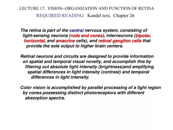
LECTURE 17: VISION--ORGANIZATION AND FUNCTION OF RETINA. REQUIRED READING: Kandel text, Chapter 26. The retina is part of the central nervous system, consisting of light-sensing neurons ( rods and cones ), interneurons ( bipolar ,

E N D
LECTURE 17: VISION--ORGANIZATION AND FUNCTION OF RETINA REQUIRED READING: Kandel text, Chapter 26 The retina is part of the central nervous system, consisting of light-sensing neurons (rods and cones), interneurons (bipolar, horizontal, and amacrine cells), and retinal ganglion cells that provide the sole output to higher brain centers. Retinal neurons and circuits are designed to provide information on spatial and temporal visual novelty, and accomplish this by filtering out absolute light intensity (brightness)and amplifying spatial differences in light intensity (contrast) and temporal differences in light intensity. Color vision is accomplished by parallel processing of a light region by cones possessing distinct photoreceptors with different absorption spectra.
Gross Anatomy of Retina Light ray
Rhodopsins: GPCRs With Tethered Light-Activated Ligand Rhodopsin consists of a GPCR called opsin, on which is linked a vitamin A derivative called retinal. When retinal is an all-trans isomer, it activates opsin, which signals to the heterotrimeric G protein transducin. 11-cis retinal All-trans retinal PHOTON
Light-Activated Rhodopsin Turns Off a Dark Current Through cGMP-gated Cation Channel
Photoreceptor Cells and Bipolar Cells Do Not Fire Action Potentials Voltage in photoreceptor cells ranges between -40 mV (when dark current is on) down to -70 mV (when current is shut off). The cGMP-gated cation channels conduct calcium as well as sodium, thereby mediating neurotransmitter glutamate release. Light-induced hyperpolarization stops glutamate release. Target bipolar cells also have cGMP-gated channels, and depolarization of bipolar cells also promotes glutamate release.
Rods and Cones Have Different Distributions Across Retina Only our central foveal vision can detect color
Cones Each Express One of Three Distinct Photoreceptors Rods All Express a Common Photoreceptor Not Found in Cones Rods can detect as little as ONE PHOTON of light, while cones are less sensitive
Basis of Color Perception Lies In Overlapping But Distinct Absorption Spectra of Cone Photoreceptors
Receptive Field Reflects Vertical and Horizontal Neural Projections: On-Center and Off-Center Bipolar Cells Any cone signals vertically to both off-center and on-center bipolar cell. Rod cells only signal through on-center bipolar cells. http://webvision.med.utah.edu/book/part-v-phototransduction-in-rods-and-cones/bipolar-cell-pathways-in-the-vertebrate-retina/ Off-Center Bipolar Has elevated resting potential Hyperpolarizes when cone is stimulated Has ionotropic GluRs On-Center Bipolar Depolarizes when cone is stimulated Has metabotropic GluRs that hyperpolarize cell (in some cases, by same mechanism as rhodopsin)
Off-Center Pathway Causes Ganglion Cells to Stop Firing In Response to Light Center and Dark Surround
Horizontal Cells Provide Lateral Inhibition That Enhances Detection of Contrast
Functional Reasons for Parallel ON and OFF Bipolar Cells ON bipolar cells are more responsive to dim light than are OFF bipolar cells. Therefore, loss of ON bipolar cell function causes night blindness. Possibly because of their different sensitivities to light intensities, the precise boundaries of light contrast detection are somewhat different. A computer emulation of “edge detection” using retinal receptive fields. ON-center and OFF-center stimulation is shown in red and green respectively.