Download
1 / 19
260 likes | 1.38k Views
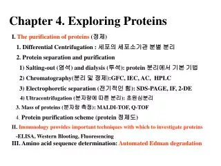
5. SEPARATION AND DETECTION OF PROTEINS II SDS-PAGE Jana Vobořilová, Anna Kotrbová-Kozak, Vlasta Fürstová, Tereza Kopská. SDS-PAGE ( = sodium dodecylsu lph ate-polyacrylamide gel electrophoresis) -met hod for separation of protein s according to their molecular weight.

E N D
5. SEPARATION AND DETECTION OF PROTEINS IISDS-PAGEJana Vobořilová,Anna Kotrbová-Kozak,Vlasta Fürstová,Tereza Kopská
SDS-PAGE(= sodium dodecylsulphate-polyacrylamide gel electrophoresis)-methodfor separation of proteinsaccording to their molecular weight
Outline of second part of the experiment*Prepare polyacrylamide gels*Add diluted samples to the samplebuffer*Heat to 95C for 4 minutes*Load the samples onto polyacrylamide gel*Run 200 volts for 30-40 minutes*Stain in Coomassie Blue stain*Destain*Identify molecular markers, actin and myosin in the separated proteins
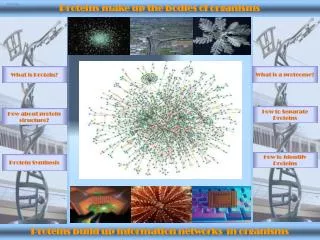
Levels of Protein Organization Primary structure = linear chain of amino acids • Secondary structure = domains of repeating structures, such as β-pleated sheets and α-helices • Tertiary structure = 3-dimensional shape of a folded polypeptide, maintained by disulfide bonds, electrostatic interactions, hydrophobic effects • Quaternary structure = several polypeptide chains associated together to form a functional protein
-proteins denatured by heating them in a sample buffer containing sodium dodecyl sulphate(SDS)-the proteins no longer have any secondary, tertiary or quaternary structure
-resultant proteins take on a rod-like shape and a uniform negative charge-to-mass ratio proportional to their molecular weights
Migration of such proteins in electric field:-negatively charged proteins move towards the positive pole-migration of proteins: *directly proportional to the overall charge of proteins *inversely proportional to protein size (molecularweight)
s-s SDS, heat - + proteins with SDS How does an SDS-PAGE gel work? • Negatively charged proteins move to positive electrode • Smaller proteins move faster • Proteins separate by size
What is in the Sample Buffer?*Tris buffer to provide appropriate pH*SDS (sodium dodecyl sulphate)detergent to dissolve proteins and give them a negative charge*Glycerol to make samples sink into wells*Bromophenol Blue dye to visualize samples
CH3 CH2 CH2 CH2 CH2 CH2 CH2 CH2 CH2 CH2 CH2 CH2 O O O S - O SDS-Polyacrylamide Gel Electrophoresis (SDS-PAGE) • SDS (Sodium Dodecyl Sulfate) detergent • solubilizes and denatures proteins • negative charge to proteins • Heat denatures proteins SDS
Why Use Acrylamide Gels to Separate Proteins? • Acrylamide gel: tight matrix • Ideal for protein separation • Smaller pore size than agarose • Proteins much smaller than intact chromosonal DNA • average amino acid = 110 Da
Protein Size • Size measured in daltons (Da) or kilodaltons (kDa) • Dalton = atomic mass unit = corresponds to mass of hydrogen molecule (1.66 x 10 -24 gram) = defined also as 1/16 of the mass of an atom of oxygen • Average amino acid = 110 Da Average nucleotide pair = 649 Da
Gel Analysis Lane 1. Kaleidoscope Markers 2. Shark 3. Salmon 4. Trout 5. Catfish 6. Sturgeon 7. Actin and Myosin Standard
Muscle Contains Proteins of Many Sizes ProteinkDaFunction titin 3000 center myosin in sarcomere dystrophin 400 anchoring to plasma membrane filamin 270 cross-link filaments into gel myosin heavy chain 210slide filaments spectrin 265 attach filaments to plasma membrane nebulin 107 regulate actin assembly a-actinin 100 bundle filaments gelosin 90 fragment filaments fimbrin 68 bundle filaments actin42form filaments tropomyosin 35 strengthen filaments myosin light chain 27slide filaments troponin (T, I, C) 30, 19, 17 mediate regulation of contraction thymosin 5 sequester actin monomers
Extension of the study WESTERN Blot Analysis *transfer of separated proteins from the gel onto a membrane *identification of a protein by a complex of primary and secondary antibodies *visualization by color reaction or by chemiluminiscence
WESTERN blot(method for detection of protein):-its name is a pun off the name Southern blot, a technique for DNA detection developed earlier by Edward Southern-similarly is named Northern blot, method for detection of RNA