Download
1 / 79
810 likes | 1.1k Views
CBP: Cardiac Arrest. Case Presentation. A 55 year old business man collapses at work. This is witnessed by his colleagues who find him pulseless . They initiate CPR and call 911.

E N D
Case Presentation A 55 year old business man collapses at work. This is witnessed by his colleagues who find him pulseless. They initiate CPR and call 911. • EMS arrive 5 minutes later. They confirm the pulseless state and place the patient on a monitor; he is in V. Fib. Standard ACLS protocols are initiated; the patient is intubated and transported to the closest ED. • The patient arrives at the ED 7 minutes later. He has received 2 doses of Epinephrine and one dose of Atropine. He has received 2 shocks and is currently in PEA arrest.
In the ERP confirms ETT placement, the rhythm of PEA, and performs a quick bedside ECHO, all the while continuing with CPR. The ECHO shows cardiac motion. • The patient is given another dose of Epinephrine and Atropine. By 6 minutes of his arrival, he is noted to have Return of Spontaneous Circulation and to have reverted to NSR. • ICU is consulted
Vital signs: HR – 112, RR – 6/poor effort, BP 65/40 (MAP 48), 36.5 Rectal Temp, Glucose 17.8, Sat’n 100%. • Quick exam reveals: A: ETT in place. B: GBS x2. +ve ETCO2 Capnography. C: As above. N HS. D: GCS of 3T, absent gag/corneal/papillary response. E: Nothing obvious. And no calf edema. • Past medical history reveals a 30 pack-year smoking history. He is on no meds and has no known drug allergies. He is known to travel abroad frequently with his work.
Question 1 • Please define Post-Cardiac Arrest Syndrome and its 4 pathophysiologic components. (Erik)
Definition • Post-cardiac arrest syndrome is a unique and complex combination of pathophysiological processes, which include • post-cardiac arrest brain injury, • post-cardiac arrest myocardial dysfunction, and • systemic ischemia/reperfusion response. • This state is often complicated by a fourth component: 4. the unresolved pathological process that caused the cardiac arrest.
Phases – for Therapy & for Science • The immediate post-arrest phase could be defined as the first 20 minutes after ROSC. • The early post-arrest phase could be defined as the period between 20 minutes and 6 to 12 hours after ROSC, when early interventions might be most effective. • An intermediate phase might be between 6 to 12 hours and 72 hours, when injury pathways are still active and aggressive treatment is typically instituted. • Finally, a period beyond 3 days could be considered the recovery phase, when prognostication becomes more reliable and ultimate outcomes are more predictable.
Pathophysiology • The 4 key components of post-cardiac arrest syndrome are: • post-cardiac arrest brain injury, • post-cardiac arrest myocardial dysfunction, • systemic ischemia/reperfusion response, and • persistent precipitating pathology.
Pathophysiology • The unique features of post-cardiac arrest pathophysiology are often superimposed on the disease or injury that caused the cardiac arrest, as well as underlying comorbidities. • Therapies that focus on individual organs may compromise other injured organ systems. • The severity of these disorders after ROSC is not uniform and will vary in individual patients based on the severity of the ischemic insult, the cause of cardiac arrest, and the patient’s pre-arrest state of health.
Foundation on which to grow… • In a study of dogs with induced cardiac arrest… • In a single observational human study… • Biochemical and neurohormonal models suggest… • A growing body of evidence… • These findings suggest, in theory, that… • These findings do not rule out the potential effect of… • Limited evidence is available to guide…
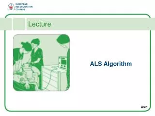
Fundamentals • Who remembers 5:1, 15:2, 30:2, 10:1, vs continuous? (AHA, ACC, ILCOR) • ETT vs supraglottic device? (AHA, ACC, ILCOR) • BLS plus AED vs ACLS (OPALS, PAD) • Push hard, push fast, push often! (ROC-BC)
Question 2 • How do you treat Post-Cardiac Arrest Syndrome. (Federico)
Early HD optimization • No evidence based guidelines • Suggestion is to have a similar approach as EGDT for Sepsis • MAP goals undefined • Loss of Cerebral Autoregulation • CPP dependent on MAP • ICP generally not elevated
MAP Goals >65, <90 • Mixed venous gases • Venous Hyperoxia • Falsely elevated levels due to poor tissue extraction related to epi use and mitochondrial failure • Follow urine output (careful in hypothermia) • Follow lactates (need to follow trends)
Avoid hyperoxia • Ptl for increased free radical production • PaO2 goals of 92 – 96% • Aim for normocarbia • Volume resuscitate • Consider intropes/vasopressors
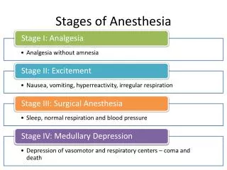
Treat for ACS • Noemie • Hypothermia • Ibrahim • Treat seizures • Increase cerebral metabolism • No Evidence for prophylaxis • Myoclonus • Clonazepam
Treat hyperglycemia • No evidence for Neuroprotective medications • Adrenal dysfunction • Renal failure • Infection • More prone to aspiration pneumonia
Question 3 • Should we cool this patient? Who do we cool, what parameters do we use, what are the complications of hypothermia therapy? What if the original documented rhythm was PEA? (Ibrahim)
Who should be cooled? • Out-of-hospital VF/PVT Arrest • RCTs:HACA and Bernard et al (NEJM, 2002): significant survival to d/c and neuro recovery (NNT 6 in a meta-analysis) • In-hospital VF Arrests • Small subset within HACA: favorable survival • Out-of-hospital all-rhythms, or non-VF • 4 retrospective studies for all (Oddo 2006, Scott 2006, Arrich 2007, Hay 2008), one retrospective and 2 observational for non-VF: possible benefit • Pyrexia within 72 Hr (>37C--> poor neuro outcomes), all patients
What are the parameters of cooling protocol? • Target core temp: 33C, or 32-34C • Onset: variable, ASAP (2-8 Hr, up to 24Hr) • Duration: 12-24 Hr • Further data required • NRCPR, HACA-R
Complications of TH • Technical: Shivering, use of ongoing sedation and NMB, to prevent shivering (with 30% dec clearance with T=34C), fluctuations of temp • HD: inc SVR, dec COP, arrhythmias (esp brady) • Diuresis, hypovolemia, dec K, Ca, Mg, PO4 --> arrhythmia • MgSO4: NMDA blocker, so dec shivering, vasodilator, so facilitate cooling induction, antiarrhythmic, and ? additive Neuroprotective (animal data)
Impaired glucose tolerance (dec insulin level and sensitivity) • Coagulopathy • Lower immunity--> infections • Higher pneumonias in TH group in HACA, but NS
Should we cool this patient? • Yes! Out-of-hospital VF arrest
Question 4 • His wife has just arrived with his 3 kids (16, 15, and 9 years old). They want to know what his prognosis is. What do you tell them and how do you prognosticate patients post arrest? Please discuss clinical and lab findings and imaging modalities. Would things be looked at differently if he was cooled? (Neil)
. What do you tell them and how do you prognosticate patients post arrest? • Timing • What is a “poor outcome”? • Prognostication • Clinical • EEG • Biomarkers • Imaging
Timing • Very difficult to prognosticate in the first 24 hours • Most evidence is derived on testing at 72 hours • Therapeutic hypothermia changes the timeline
What is a poor outcome? • Poor outcome is defined as death, unconsciousness after one month, or unconsciousness or severe disability after six months.
Clinical signs • Absence of pupillary light reflexes • 100% specificity in meta analysis • LR+ 10.5 (CI 2.1-52.4) • Absence of motor response to pain • 100% specificity in meta analysis • LR+ 16.8 (CI 3.4 – 84.1) • Myoclonic status epilepticus • Can be predictive early • Much worse than SE
Clinical Signs • Which are not good prognositcators • Age • Sex • Cause of arrest • Type of arrhythmia • Total arrest time • Duration of CPR
EEG • Overall prognostication ability is not strong • Variety of studies have looked into it • Lack of a standardized classification system • Concerning features • Burst suppression • Nonreactive alpha and theta patterns • Generalized periodic complexes
SSEP’s • Tests integrity of the neuronal pathways from peripheral nerve, spinal cord, brainstem, and cerebral cortex
N20 • Best studied waveform • Robust as it is not strongly influence by meds and metabolic derangements • LR+ 12 (CI 5.3-27.6)
Biomarkers • Dead brain releases biomarkers • 3 have been “well” studied • Neuron specific enolase (NSE) • S-100 • Creatinine kinase BB isoenzyme (CK-BB)
Imaging • Although not strong enough to prognosticate reliably, a bad scan is a bad scan • Problem lies in that a good scan may not be a good scan
Question 5 • His EKG shows normal sinus rhythm with non-specific changes. Should he go to the cath lab? If so, what are the recommendations for cath post cardiac arrest? If he arrested again, would you thrombolyse him? What is the etiology of the vast majority of cardiac arrests? (Noamie)
Questions • His EKG shows normal sinus rhythm with non-specific changes. Should he go to the cath lab? • If so, what are the recommendations for cath post cardiac arrest? • If he arrested again, would you thrombolyse him? • What is the etiology of the vast majority of cardiac arrests?
65-70 % 10% 5-10% 15 to 35%
Etiology of Sudden Cardiac Death • Age < 20: • Myocarditis (22%), HCM (22%) and conduction system abnormalities (13%) • Age 20-29: • CAD (24%), myocarditis (22%) and HCM (13%). • Age 29-39: • CAD (58%), myocarditis (11%). Am J Cardiol 1991;689(13):1388-1392
Should he go to the cath lab? • Yes • Even if no evidence of an ACS, need to exclude stable/chronic CAD • Sudden cardiac arrest may be first indication of CAD • But, does he need it right now?