Download

1 / 63
770 likes | 1.75k Views
Clinical Correlate: Examination of Nystagmus. Najwa Al-Bustani Neurology AHD July 20-2011. Objectives:. Define Nystagmus. How to describe Nystagmus. Types. Physiological & Physilogical Nystagmus. What is Nystagmus ?.

E N D
Clinical Correlate:Examination of Nystagmus Najwa Al-Bustani Neurology AHD July 20-2011
Objectives: • Define Nystagmus. • How to describe Nystagmus. • Types. • Physiological & Physilogical Nystagmus.
What is Nystagmus ? • A repetitive rhythmic involuntary oscillation of the eyes that is usually conjugate. • Biphasic ocular oscillation containing slow eye movement that are responsible for its genesis and continuation.
Waveforms: • Jerk (most common): slow drift ‘slow phase’ followed by quick reset ‘quick phase’. • Slow phase waveforms can be: • Increasing velocity exponential = congenital. • Decreasing velocity exponential = gaze-evoked. • Constant (linear) velocity = vestibular. 2. Pendular: sinusoidal oscillation ‘like pendulum’, phases have equal speed.
Trajectory: • Horizontal. • Vertical. • Torsional. • Combination of all three.
Direction: • Usually defined by its fast phase. Conjugacy: • Conjugate: both eyes move in same direction. • Disconjugate: eyes move in different direction >> disjunctive.
Alexander’s law: • Jerk nystagmus usually increases in intensity when looking in the direction of the fast phase. Null zone: • The field of gaze where nystagmus intensity is minimal.
Grading of jerk Nystagmus: • Grade 1: present only when looking in the direction of the quick component. • Grade 2 : also present when looking straight ahead. • Grade 3 : present when looking in the direction of the quick component, when looking straight ahead and when looking in the direction of the slow component.
Physiologic Nystagmus: • End-position: few beats of horizontal nystagmus when the eyes are first moved to extreme horizontal positions in the orbit.
Vestibular Nystagmus: • Caloric Nystagmus: • Irrigation with worm water (in the head up supine position) causes endolymph in the horizontal canal to move toward the ampulla, exciting the hair cells & driving a slow phase eye movement away from the irrigated side. • Cold water: inhibits the horizontal canal, produce slow phase toward the irrigated side.
Vestibular Nystagmus: • Rotational Nystagmus (VOR): • Prolonged head rotation produces a slow phase in the direction opposite to the head movement interrupted by quick phases in the same direction as head movement. • This serves to stabilize retinal images as head moves.
Optokinetic Nystagmus: • Driven by prolonged full-field visual motion. • Supplements the VOR to stabilize vision.
http://www.youtube.com/watch?v=yo-zA1CuKUI&feature=related • 38 year old female, c/o vomiting X few hours. • ? left earache and tinnitis.
Peripheral Vestibular Nystagmus: • Jerk nystagmus due to imbalance of vestibular inputs.
Unilateral Vestibular hypofunction: • Acute lesion to one labyrinth or vestibular nerve >> spontaneous horizontal/torsional nystagmus. • Because of the tonic input from the intact side is suddenly unopposed.
Unilateral Vestibular hypofunction: • The eyes drift (slow phase) toward the lesioned side and quick phase beat toward the intact side. • The intensity usually greatest when looking toward the intact side (Alexander’s law). • Direction does not change with gaze, unlike gaze-evoked nystagmus.
Unilateral Vestibular hypofunction: • May be partially or fully suppressed by vision and is best seen when fixation is removed: • Frenzel goggles. • When looking at one optic nerve with direct ophthalmoscope, cover the fellow eye.
Vestibular hypofunction: • Bilateral vestibular lesions do not cause nystagmus because the lesion is symmetric and there is no imbalance.
Bruns Nystagmus Primary position Rt. Lt.
Bruns Nystagmus: • May be seen in large tumors in the CP angle. • 2 components: • Horizontal nystagmus beating away from the lesion when looking away from the lesion (vestibular nystagmus-accentuated by Alexander’s law) due to vestibular nerve involvement.
Bruns’ Nystagmus: • Horizontal nystagmus beating toward the lesion when looking toward the lesion (unilateral gaze-evoked nystagmus) due to compression of the adjacent brainstem and cerebellar flocculus.
Benign Paroxysmal Positional Nystagmus (BPPN): • Brief (<1min) nystagmus provoked by changes in head position relative to gravity. • Caused by free-moving otoconia that have become lodged in a semicircular canal. • Usually affects the posterior canal: slow phase directed downward with a torsional component in which the upper pole of the eyes rotate away from the affected ear (upbeating-torsional nystagmus).
Benign Paroxysmal Positional Nystagmus (BPPN): • Diagnosed by Dix-Hallpike maneuver. • Treated by repositioning maneuvers (Epley) that move the otoconia out of the affected canal.
Congenital Nystagmus: • May be present at birth, more commonly appears later in infancy. • Sporadic or Genetic: AD (6p12), AR, X-linked recessive. • Associated with oculocutaneous albinism. • Commonly accentuated by attempted fixation or anxiety. • Typically damped by eye closure, sleep & convergence.
Congenital Nystagmus: • Does not cause oscillopsia. • Distinct waveforms: • Conjugate horizontal-torsional pendular and/or jerk nystagmus. • Similar amplitude in both eye. • Jerk nystagmus has increasing velocity slow phases.
Congenital Nystagmus: • Foveation periods: brief cessation of eye motion, often following quick phases, during which clear vision is possible (if there are no afferent visual abnormalities). • There is often orbital position (null point) where nystagmus is minimal and vision is best.
Congenital Nystagmus: • May be associated with other abnormalitis: • Strabismus, latent nystagmus, head oscillations.
Spasmus Nutans: • Triad of: head turn, head nodding and nystagmus. • Typically develops during the first year of life. • Resolves by age 10. • Horizontal or vertical pendular nystagmus with low amplitude and high frequency.
Spasmus Nutans: • May be monocular or of different amplitude and/or phase in each eye. • Optic pathway glioma can cause acquired monocular nystagmus and should be ruled out by MRI.
Convergence-retraction nystagmus: • Convergence and/or retraction of the eyes elicited by attempted upward saccades or quick phases. • Best seen during stimulation with a downward-moving OKN stimulus.
Convergence-retraction nystagmus: • Part of the dorsal midbrain syndrome: • Impaired vertical gaze (particularly upwoard). • Light-near dissociation of pupillary responses. • Lid retraction (Collier’s sign). • Convergence-retraction nystagmus. • Spasm or paresis of convergence, accommodation. • Skew deviation.
Convergence-retraction nystagmus: • Due to lesion affecting the area of the posterior commissure: • Tumors (pineal). • Hydrocephalus (e.g aqueductal stenosis). • Hemorrhage or infarction (midbrain, thalamus). • Multiple sclerosis or inflammatory lesions.
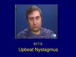
Upbeat Nystagmus: • Spontaneous nystagmus with downward slow phases in primary position. • Etiologies: • Focal lesions: (infarction, tumor, demyelinating) of the medulla or cerebellum. • Cerebellar degeneration. • Wenicke’s encephalopathy.
Downbeat Nystagmus: • Spontaneous upward drift of the eyes. • Characteristic sign of a lesion of the vestibulocerebellum or its pathway or its pathway in the brainstem (cerebellar degeneration, MS, stroke). • Other causes: drug toxicity ‘Li, AED’, Wernicke’s encephalopathy.
Downbeat Nystagmus: • Often occurs in the context of a more general cerebellar syndrome but may present in isolation & be progressive. • Slow phase waveform may have constant, decreasing, or increasing velocity. • Commonly enhanced by down and lateral gaze.
Gaze-Evoked Nystagmus: • Loss of eccentric gaze holding, eyes tend to drift back to the center of the orbit. • Weak neural intergrator does not produce suffeciently strong tonic innervation to hold the eyes against elastic forces.
Gaze-Evoked Nystagmus: • Direction of nystagmus depends on gaze direction. • Typically horizontal. • May also include upbeat nystagmus with upgaze. • With sustained gaze >> nystagmus will often diminish.
Gaze-Evoked Nystagmus: • Upon return to center position, there may be a brief oppositely directed nystagmus (rebound nystagmus). • Gaze-evoked & downbeat nystagmus often occur together in patients with cerebellar degeneration.
Gaze-Evoked Nystagmus: • Etiologies: • Vestibulocerebellar lesions >> cerebellar degeneration. • Functional neural integrator impairment due to drugs (sedatives, AED), metabolic derangements.