Download
1 / 85
850 likes | 877 Views
Explore the intricacies of specific immunity, including antigen presentation, T and B cell activation, antibody production, and primary & secondary immune responses. Learn about acquired immunity types, properties of immunity, and the immune response division. Discover the roles of T cells, B cells, antigen recognition, and MHC proteins in immune defense mechanisms.

E N D
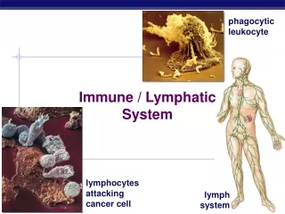
Chapters 20, 21. Lymphatic and Immune System Part II. Specific Immunity
Overview • Properties of specific immunity • Antigen presentation and the MHC complexes • T cell activation • B cell activation • Antibodies • Primary and secondary immune response • Clonal selection • Diseases
Specific Defenses • Specific resistance(immunity): • responds to specific antigens with coordinated action of T cells and B cells • Recognizes specific foreign substances • Acts to immobilize, neutralize, or destroy foreign substances • Amplifies inflammatory response and activates complement • T Cells: • provide cell-mediated immunity • defends against abnormal cells and pathogens inside cells • B cells • provide humoral or antibody-mediated immunity • defends against antigens and pathogens in body fluids
Forms of Acquired Immunity Figure 22–14
Forms of Immunity • Innate: present at birth • Acquired: after birth • Naturally acquired through normal environmental exposure • Active: antibodies develop after exposure to antigen • Passive: antibodies are transferred from mother through breastfeeding • Artificially acquired through medical intervention • Active: antibodies inducedthrough vaccines containing pathogens • Passive: antibodies injected into body
Properties of Immunity • Specificity • Each T or B cell: responds only to a specific antigen and ignores all others • Versatility • Many subtypes of lymphocytes: each fights a different type of antigen • active lymphocyte clones itself to fight specific antigen • Memory • memory cellsstay in circulation and provide immunity against new exposure • Tolerance • Immune system ignores “normal” antigens
The Immune Response Figure 22–15 (Navigator)
The Immune Response • 2 main divisions: • cell mediated immunity(T cells) • antibody mediated immunity (B cells) • MOVIE – Cell Mediated Immunity
Antigens • Substances that can mobilize the immune system and provoke an immune response • The ultimate targets of all immune responses are mostly large, complex molecules not normally found in the body (nonself)
Types of T Cells • Cytotoxic T Cells (Tc cells. CD8 cells) • Attack cells infected by viruses • Responsible for cell-mediated immunity • Helper T Cells (Th cells, CD4 cells) • Stimulate function of T cells and B cells • Suppressor T cells (Ts cells) • Inhibit function of T cells and B cells
Antigens and MHC Proteins Figure 22–16a (Navigator)
Antigen Recognition • T cells only recognize antigens that are bound to glycoproteins in cell membranes “presented” • MOVIE: antigens and MHC proteins
Antigen Presentation Figure 22–16b
“Self Antigens”: MHC Proteins • Membrane glycoproteins that differ among individuals and identify them as “self” • Bind to and present antigens • Class I: found in membranes of all nucleated cells • Class II: found in membranes of professional antigen-presenting cells (APCs): B cells, dendritic cells, macrophages
MHC Proteins • Are coded for by genes of the major histocompatibility complex (MHC) and are unique to an individual • Each MHC molecule has a deep groove that displays a peptide, which is a normal cellular product of protein recycling • In infected cells, MHC proteins bind to fragments of foreign antigens, which play a crucial role in mobilizing the immune system
Antigen Recognition • T cell receptors only recognize and bind to cells whose MHC protein contains an abnormal peptide fragment, staring an immune response • The particular antigen occupying the MHC are recognized together. • If the MHC contains a normal cellular protein fragment, T cells will not recognize it, no reaction will occur. • So T cells must simultaneously recognize: • Nonself (the antigen) • Self (a MHC protein of a body cell)
MHC Proteins • Both types of MHC proteins are important to T cell activation • Class I MHC proteins • Always recognized by CD8 T cells • Display peptides from endogenous antigens • Class II MHC proteins • Always recognized by CD4 T cells • Display peptides from external antigens
Class I MHC Proteins • These MHCs pick up small peptides from inside the cell and carry them to the surface and “present” them to Tc cells • T cells ignore normal peptides • Abnormal peptides or viral proteins activate T cells to destroy cell
Class I MHC Class II MHC
Class II MHC Proteins • Found only on professional APCs • These cells ingest external pathogens and processes them • Antigenic processing (chopping up) of pathogens brought into cells • Fragments bind to Class II proteins • MHC plus fragments are inserted into cell membrane and presented to Th cells
Antigen Presenting Cells APCs • APCs are responsible for activating T cells against foreign cells and proteins • Phagocytic • Free and fixed macrophages: • in connective tissues • Kupffer cells: • of the liver • Microglia: • in the CNS • Pinocytic • Langerhans cells: • in the skin • Dendritic cells: • in lymph nodes and spleen • B cells
CD Markers • Also called cluster of differentiation markers: found in T cell membranes • CD3 Receptor Complex– All T cells • CD8- cytotoxic T cells and suppressor T cells • CD4 - Found on helper T cells
T Cell ActivationStep 1: antigen binding • CD8 or CD4 binds to CD3 receptor complex and prepare cell for activation • CD8 helps bind to MHC Class I (cell types?) • CD4 helps bind to MHC Class II (cell types?) • APCs produce co-stimulatory molecules that are required for TC activation • Mobile APCs (Langerhans’ cells) can quickly alert the body to the presence of antigen by migrating to the lymph nodes and presenting antigen
T Cell Activation Step 2:Costimulation • For T cells to be activated, it must be costimulated by binding to a stimulating cell (APC) at second site (in addition to the TCR-MHC interaction) which confirms the first signal • This is antigen nonspecific and can be provided by: • interaction between co-stimulatory molecules expressed on the membrane of APC and the T cell (like a second lock and key) • Cytokines sent from APC to T cell • Redundancy limits errors of inappropriate activation: without co-stimulation, T cells: • Become tolerant to that antigen • Are unable to divide • Do not secrete cytokines
Activated T cells • After antigen recognition and co-stimulation, T cells: • Enlarge, proliferate, and form clones • Differentiate and perform functions according to their T cell class • Primary T cell response peaks within a week after signal exposure • Effector T cells then undergo apoptosis between days 7 and 30 • The disposal of activated effector cells is a protective mechanism for the body • Memory T cells remain and mediate secondary responses to the same antigen
Actions of Cytotoxic T Cells • Killer T cells(Tc) seek out and immediately destroy target cells (only T cells that do hand-to-hand combat) Q: What are the targets? • Release perforin to lyse target cell membrane • Secrete poisonous lymphotoxin to destroy target cell • Activate genes in target cell that cause cell to die • Create Memory Tc Cells which stay in circulation and immediately form cytotoxic T cells if same antigen appears again
Activation of Cytotoxic T Cells Figure 22–17 (Navigator)
2 Classes of CD8 T Cells • Activated by exposure to antigens on MHC proteins: • one responds quickly: • producing cytotoxic T cells and memory T cells • the other responds slowly: • producing suppressor T cells • Suppressor T Cells • Secrete suppression factors • Inhibit responses of T and B cells • After initial immune response • Limit immune reaction to single stimulus
Helper T Cells (Th) • Once primed by APC presentation of antigen, Activated CD4 T cells divide into • effector Th cells, which secrete cytokines • memory Th cells, which remain in reserve • Effectors chemically or directly stimulate proliferation of other T cells and stimulate B cells that have already become bound to antigen • Without TH, there is no immune response
Activation of Helper T Cells Figure 22–18
Functions of T Cell Cytokines • Stimulate T cell divisions: • produce memory T cells • accelerate cytotoxic T cell maturation • Attract and stimulate macrophages • Attract and stimulate NK cells • Promote activation of B cells
Th cells are required for B cells to become active Th cells help Tc cells become active
Pathways of T Cell Activation Figure 22–19
Importance of Cellular Response • T cells recognize and respond only to processed fragments of antigen displayed on the surface of body cells • T cells are best suited for cell-to-cell interactions, and target: • Cells infected with viruses, bacteria, or intracellular parasites • Abnormal or cancerous cells • Cells of infused or transplanted foreign tissue
Complete immune response
B Cells • Responsible for antibody-mediated immunity • Attack antigens by producing specific antibodies • Millions of populations, each with different antibody molecules • MOVIE: Antibody mediated immunity
Step 1:B Cell Sensitization • Corresponding antigens in interstitial fluids bind to B cell receptors (which are antibodies • B cell prepares for activation, a process called sensitization • MOVIE B cell sensitization
B Cell Sensitization • During sensitization, antigens are taken into the B cell along with surface receptor (antibody), processed, and then reappear on surface, bound to Class II MHC protein • Remember, B Cells are one the of “the professional APCs” (any cell with MHC Class II is an APC) • Sensitized B cell is prepared for activation but does NOT yet divide; it needs stimulation by a helper T cell that has been activated by the same antigen
Step 2: B Cell Activation • A specific Helper T cell binds to MHC Class II complex on the sensitized B cell which contains peptide fragment • Th secretes cytokines that promote B cell activation and division • Activated B cell divides into: • plasma cells:synthesize and secrete antibodies into interstitial fluid • memory B cells: like memory T cells remain reserve in the body to respond to next infection immediately
B Cell Sensitization and Activation Figure 22–20 (Navigator)
Adaptive Immunity: Summary • Two-fisted defensive system that uses lymphocytes, APCs, and specific molecules to identify and destroy nonself particles • Its response depends upon the ability of its cells to: • Recognize foreign substances (antigens) by binding to them • Communicate with one another so that the whole system mounts a response specific to those antigens
Antibodies (Immunoglobins) • Soluble proteins secreted by activated B cells and plasma cells in response to an antigen • Found in body fluids • Capable of binding specifically with that antigen • Structure • 2 parallel pairs of polypeptide chains: • 1 pair of heavy chains • 1 pair of light chains • Each chain contains: • constant segments: determine the type of antibody (IgG, IgE, IgD, IgM, IgA) • variable segments: determine specificity of the antibody • Free tips of the 2 variable segments form antigen binding sites of antibody molecule
Antibody Structure Figure 22–21a, b
5 Classes of Antibodies • Class is determined by constant segments • Class has no effect on antibody specificity • IgG: (80%). Most important class, cross placenta providing passive immunity to fetus • IgE: help against worms and parasites; activate basophils and mast cells to release histamine allergy • IgD: Important in B cell acivation • IgM: Pentameric. First class to be secreted during primary response. • IgA: found in mucosal secretions, glands, epithelia (including breast milk)
Antibody Function • Antibodies themselves do not destroy antigen; they inactivate and tag it for destruction • All antibodies form an antigen-antibody (immune) complex • Defensive mechanisms used by antibodies are neutralization, agglutination, precipitation, and complement fixation Figure 22–21c, d
Functions ofAntigen–Antibody Complexes • Antigen–Antibody Complex = an antibody bound to an antigen. Causes: • Neutralization of antigen binding sites • Precipitation and agglutination: formation of immune complex • Activation of complement (main mechanism against cellular antigens) • Opsonization: increasing phagocyte efficiency • Stimulation of inflammation
Antibody Function - Summary • Antibodies produced by active plasma cells bind to target antigen and: • inhibit its activity • destroy it • remove it from solution • promote its phagocytosis by other defense cells • Importance of humoral response: • Soluble antibodies are the simplest ammunition of the immune response • Interact in extracellular environments such as body secretions, tissue fluid, blood, and lymph