Download
1 / 57
610 likes | 1.09k Views
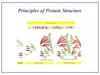
Secondary structure of proteins : turns and helices. Levels of protein structure organization. Peptide bond geometry. Hybrid of two canonical structures. 60% 40%. Electronic structure of peptide bond. Peptide bond: planarity.

E N D
Peptide bond geometry Hybrid of two canonical structures 60% 40%
Peptide bond: planarity • The partially double character of the peptide bond results in • planarity of peptide groups • their relatively large dipole moment
Side chain conformations: the c angles c1 c2 c3 c1=0
Dihedrals with which to describe polypeptide geometry side chain main chain
Peptide group: cis-trans isomerization Skan z wykresem energii
Because of peptide group planarity, main chain conformation is effectively defined by the f and y angles.
The dihedral angles with which to describe the geometry of disulfide bridges
Some andpairs are not allowed due to steric overlap (e.g, ==0o)
Conformations of a terminally-blocked amino-acid residue E Zimmerman, Pottle, Nemethy, Scheraga, Macromolecules, 10, 1-9 (1977) C7eq C7ax
Energy maps of Ac-Ala-NHMe and Ac-Gly-AHMe obtained with the ECEPP/2 force field
Energy curve of Ac-Pro-NHMe obtained with the ECEPP/2 force field fL-Pro»-68o
Energy minima of therminally-blocked alanine with the ECEPP/2 force field
Elements of protein secondary structure • Turns (local) • Loops (local) • Helices (periodic) • Sheets (periodic) • Statistical coil (not regular)
g- and b-turns g-turn (fi+1=-79o, yi+1=69o) b-turns
Types of b-turns in proteins Hutchinson and Thornton, Protein Sci., 3, 2207-2216 (1994)
Older classification Lewis, Momany, Scheraga, Biochim. Biophys. Acta, 303, 211-229 (1973)
fi+1=-60o, yi+1=-30o, fi+2=-90o, yi+2=0o fi+1=60o, yi+1=30o, fi+2=90o, yi+2=0o fi+1=-60o, yi+1=-30o, fi+2=-60o, yi+2=-30o fi+1=60o, yi+1=30o, fi+2=60o, yi+2=30o
fi+1=-60o, yi+1=120o, fi+2=80o, yi+1=0o fi+1=60o, yi+1=-120o, fi+2=-80o, yi+1=0o
cis-proline |yi+1|»80o, |fi+2|<60o |yi+1|»60o, |fi+2|»180o
Helical structures a-helical structure predicted by L. Pauling; the name was given after classification of X-ray diagrams. Helices do have handedness.
Geometrical parameters of helices Average parameters of helical structures Turns closed by H-bond H-bond radius Type
Idealized hydrogen-bonded helical structures: 310-helix (left), a-helix (middle), p-helix (right)
a-helices: deformationsbifurcated or mismatched H-bonds disrupt periodic structure 1,6-hydrogen bonds at helix ends. Bifurcated hydrogen bonds (1,4 and 1,5) at helix ends. 1,3-, and 1,4-hydrogen bonds at helix ends.
Bifurcated hydrogen bonds (1,4 and 1,5) at the N-terminums of helix A of thermolysin. Zniekształcenia a-helisdodatkowe wiązania wodorowe na końcachhelis (wiązanie wodorowe rozwidlonelub zmiana wiązania wodorowego) Bifurcated hydrogen bonds (1,4 and 1,5) at the C-terminums of helix D of carboxypeptidase. 1,6 and 2,5 hydrogen bonds at the C-terminus of helix A in lysosyme
Helix deformation (kink) Example from myoglobin structure. The kink angle is up to 20o
Other factors resulting in helix deformation • Deformation is forced because of tertiary structure (crowding). • Strong H-bonding (e.g., between side chains). • Helix breakers inside; Pro will result in a kink for sure and Gly almost always but small polar amino acids such as Ser and Thr also can. Kink inside an a-helix in phosphoglyceryl aldehyde dehydrogenase
Helix breaking by Pro residues Ring constraint No amide hydrogen O C-O N H
Helix capping The first and the last residue are the capping residues The N1 and C1 residues possess f and y angle values typical of an a-helix About 48% residues in Ncap-N1-N2-N3 fragments and about 35% of residues in -C3-C2-C1-Ccap- fragments forms hydrogen bonds in which side-chain groups take part. ...-N’’-N’-Ncap-N1-N2-N3-...........................-C3-C2-C1-Ccap-C’-C’’-... Izolowana 12-resztowa a-helisa posiada 12 grup donorowych NH oraz 12 grup akceptorowych CO wiązania wodorowego (w obrębie łańcucha głównego). W 12 resztowej helisie może utworzyć się tylko 8 wewnątrzcząsteczkowych wiązań wodorowych. N- i C-Końcowy fragment helisy zawiera więc 4 wolne donory NH i 4 wolne akceptory CO wiązań wodorowych. Kompensacją tej niedogodności jest występowanie polarnych reszt aa na N- i C-końcu helisy. N- i C-Końcowe fragmenty helis wykazują dodatkowo różne preferencje co do określonych reszt aa. Residue preferences to occur at end or close-to-end positions
Contact interactions occur between the side chains separated by 3 residues in amino-acid sequence
Schematic representation a-helices: helical wheel 3.6 residues per turn = a residue every 100o.
Amphipatic (or amphiphilic) helices One side contains hydrophobic amino-acids, the other one hydrophilic ones. In globular proteins, the hydrophilic side is exposed to the solvent and the hydrophobic side is packed against the inside of the globule Hydrophobic Hydrophilic Amphipatic helices often interact with lipid membranes hydrophilic head group aliphatic carbon chain lipid bilayer
Length of a-helices in proteins 10-17 amino acids on average (3-5 turns); however much longer helices occur in muscle proteins (myosin, actin)
Proline helices (without H-bonds) Polyproline helices I, II, and III (PI, PII, and PIII): contain proline and glycine residues and are left-handed. PII is the building block of collagen; has also been postulated as the conformation of polypeptide chains at initial folding stages.
Polyproline ring conformations C2 (half-chair) conformations of Cg-endo L-proline CS (envelope) conformation of Cg-endo L-proline peptide group at the trans position with respect to Ca-H (Y=120o), as in collagene CS (envelope) of Cg-egzo L-proline with the peptide group at the cis’ orientation with respect to Ca-H (Y=-60o)
Structure F Y w turns/residue residues/turn a-helix -57 -47 180 +3.6 1.5 310-helix -49 -26 180 +3.0 2.0 p-helix -57 -70 180 +4.4 1.15 Polyproline I -83 +158 0 +3.33 1.9 Polyproline II -78 +149 180 -3.0 3.12 Polyproline III -80 +150 180 +3.0 3.1 f and y angles of regular and polyproline helices