Download
1 / 47
470 likes | 628 Views
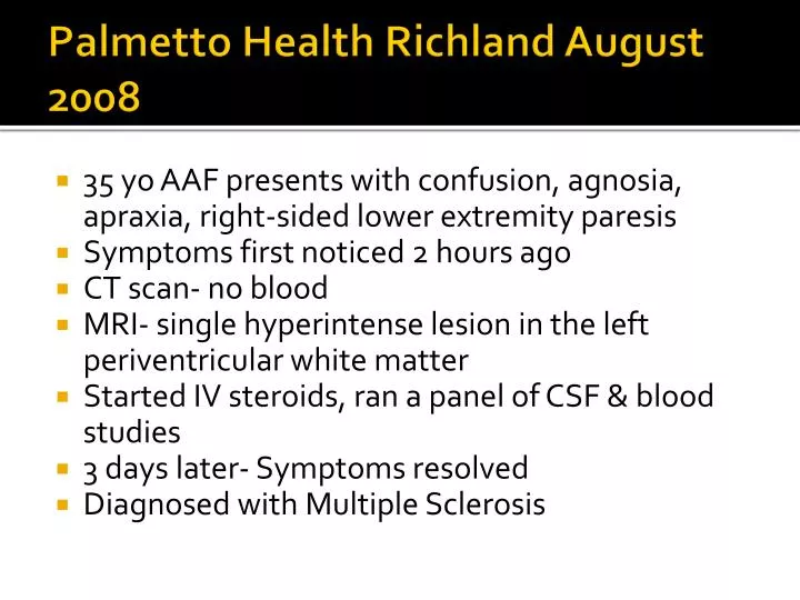
Palmetto Health Richland August 2008. 35 yo AAF presents with confusion, agnosia , apraxia , right-sided lower extremity paresis Symptoms first noticed 2 hours ago CT scan- no blood MRI- single hyperintense lesion in the left periventricular white matter

E N D
Palmetto Health Richland August 2008 • 35 yo AAF presents with confusion, agnosia, apraxia, right-sided lower extremity paresis • Symptoms first noticed 2 hours ago • CT scan- no blood • MRI- single hyperintense lesion in the left periventricular white matter • Started IV steroids, ran a panel of CSF & blood studies • 3 days later- Symptoms resolved • Diagnosed with Multiple Sclerosis
Jessica Floyd, M4 The Imaging of Multiple Sclerosis* Utility of MRI * Differential of White Matter Lesions* Future direction of neuroradiology
What is Multiple Sclerosis? • Chronic Inflammatory demyelinating disease of the CNS • 2nd-3rd decade of life (“belongs to the climax of life”) • 2:1 Female predominance • 250-350,000 people with MS in the US • Cyclical inflammatory reactions followed by remission of symptoms and variable recovery • Relapsing-Remitting- 80% • Primary Progressive- 20%; closer incidence M:F • Secondary Progressive
Charcot’s description • First described by Charcot in 1835 • Patient history, physical exam, autopsy • Salpetriere (1865) to the United States • Blood vessel at the center of each lesion • Preserved axis cyllinder • Atrophy of the medullary sheath • Types: • Cephalic • Spinal • Mixed: cerebrospinal
Broad Symptom Complex • Sensory disturbances • Unilateral optic neuritis • Diplopia- Internuclearopthalmoplegia • Nystagmus • Lhermitte’s sign • Limb weakness • Clumsiness • Gait ataxia • Neurogenic bladder • Bowel symptoms
Symptoms • Fatigue • Worse in the afternoon • Physiologic increases in temperature • Post Partum worsening of Symptoms ~ 4wk • Uhthoff’s symptom- hot shower, hot bath • Pseudoexacerbations with fever
Symptoms • Highly suggestive of MS: • Paroxysmal pain, paresthesias • Trigeminal neuralgia • Episodic clumsiness, nysarthria • Tonic limb posturing • Less common: • Prominent cortical signs • Aphasia, apraxia, recurrent seizures, visual field loss, early dementia • Extrapyramidal phenomena • Chorea, rigidity
Brain Lesions • Most sensitive modality is MRI • Sensitive to inflammation • Sensitive to demyelination • CT is a poor tool unless very severe destruction • Callosal atrophy • Whole brain atrophy
T2 Lesions • Inflammation (water) & Demyelination (loss of fat) Hyperintensities on T2 weighted images • Confirm with FLAIR images • Round, Ovoid • Vary in size. Few mm Few cm • Periventricular region, corpus callosum • Perivascular distribution, penetrating venules • Dawson’s fingers • Juxtacortical Lesions, U-fibers
T2 Lesions • Temporal Lobe • Brainstem- peripherally • Deep Gray Matter- BG, Thalamus (LC) • Cerebellum • Spinal Cord • Recurrent Lesions in Same Area CONFLUENT lesions • MC anterior & posterior to lateral ventricle • Vasogenic edema = “fuzzy extension” of T2 signal • LARGE DIFFERENTIAL FOR T2 Hyperintensities
T1 Holes • SEVERE Tissue Injury T1 dark signals • Rarely seen in the spinal cord or post fossa • Stronger correlation with demyelination & axonal loss than T2 hyperintensities • Evolution of enhancing lesions T1 Holes associated with more progressive disease
Gadolinium-Enhancing Lesions • Indicates breakdown of the blood-brain barrier • Very active inflammation • Pattern of enhancement • Homogenous • Ring reactivation of an old lesion • Heterogeneous • Enhancement duration varies- days, weeks • 5% pts have >3 months of single lesion enhancement
Spinal Cord Lesions • Round, Ovoid on T2 • Limited to 1-2 spinal cord segments • 80% involve half of cord cross sectional area • Ddx- ITM, Devic’sDz • Typically unilateral • Inflammatory edema temporary cord expansion • Ddx- Tumor (bx) • Gadolinumenhancment with active BBBB • Post mortem path studies show greater demyelination than assumed with conventional t2 imaging
Brain Atrophy • Significant Clinical Implications • Correlates with clinical disability • Predictive of later progressive disability • Many standard therapies slow progression of atrophy over time
Diagnosis • Ensuring MS is of high suspicion, consider prevalence and a priori probability
How suspicious are you? • Imaging is only one part of the story, clinical picture • Incidental Finding versus Manifesting Clinically • Normal Aging or Virchow Robin Spaces • Vascular disease • Infarction • Multi-infarct Dementia • Hypertensive encephalopathy • Sarcoidosis- ACE level, pulmonary Sx, CXR • SLE- discoid/malar rash, other organ involvment • Lyme Disease- CN7 palsy, rash, influenza-like illness • HIV- test, immunocompromised • Progressive Multifocal Leukoencephalopathy- immunocompromised • Largest differential concerns Vascular versus MS
Vascular disease vs Multiple Sclerosis 66 yo Male T2 Hyperintensities None being Ovoid Few Periventricular Lesions No Juxtacortical lesions
Criteria for Diagnosis of MS • Since MRI revolutionized the diagnosis of MS, needed specific criteria • Crux of the Dx is demonstrating attacks of neurologic dysfunction are separated in space and time • Clinical criteria* pt hx, PE findings, • Laboratory Criteria* oligoclonal bands, IgG index • MRI * 2001 McDonald Criteria, 2005 revised
One episode, treat or not to treat? • Cannot diagnose MS on MRI alone- need the clinical exam & history • However, MRI can now show us what even a vigorous clinical exam cannot • Revolutionizing treatment treat earlier • Mild cognitive deficits discovered earlier
Coming in the future… • MR Spectroscopy- N-acetyl aspartate, Lactate • Diffusion Tensor Imaging • Able to pick up on lesions not yet detectable on MRI • Ability to give you information on precisely how damaged the lesion is compared to other lesions
Diffusion Tensor Imaging • 3-D water diffusion • Mean Diffusivity- overall diffusion • Fractional anisotropy- amount of elongatedness of diffusion • Colorized primary eigenvector maps- illustrate different directions of the primary fiber tract • RED = L-R • GREEN = Up-Down • BLUE = In-Out of page