Download
1 / 38
830 likes | 2.51k Views
Parastomal hernia. Dr Chan Wai Hei, Arthur Queen Elizabth Hospital. Overview. Background Classification Risk factors Clinical presentation & Complications requiring surgical intervention Management Prevention. Definition.

E N D
Parastomal hernia Dr Chan Wai Hei, Arthur Queen Elizabth Hospital
Overview • Background • Classification • Risk factors • Clinical presentation & Complications requiring surgical intervention • Management • Prevention
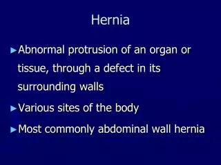
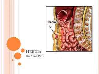
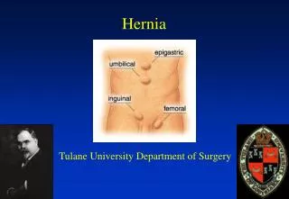
Definition • A parastomal hernia (PSH) is a type of incisional hernia that occurs at the site of stoma or immediately adjacent to the stoma • The most common late complication of a permanent stoma
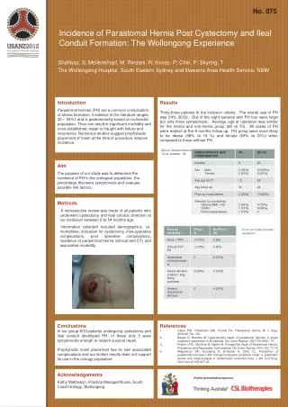
Incidence • Variable incidence reported in literature • Incidence increases with time • Most occur within 2 years of stoma formation • Some believe that it is an inevitable consequence of stoma formation
Incidence [Pilgrim CH, McIntyre R, Bailey M. Prospective audit of parastomal hernia: prevalence and associated comorbidities. Dis Colon Rectum 2010;53:71-6]
Incidence • Literature review by Carne et al. • 1.8-28.3% in end ileostomies • 0-6.2% in loop ileostomies • 4.0-48.1% in end colostomies • 0-30.8% in loop colostomies [Carne PW, Robertson GM, Frizelle FA. Parastomal hernia. Br J Surg 2003;90:784-93]
Classification • Traditional • Radiological
Classification - Traditional • 4 subtypes • 1) Subcutaneous • most common type • the herniation enters into the subcutaneous fat alongside the stoma • 2) Interstitial • the herniation extrudes alongside the bowel for stoma, then burrows into one of the intermuscular planes • 3) Peristomal • the stomal bowel is prolapsed and loops of bowel and/or omentum enter the hernia space produced between the layers of prolapsed bowel • 4) Intrastomal • enters the plane between the merging and the everted part of bowel • usually occurs in the spout type of stoma – e.g. ileostomy [Devlin HB. Peristomal hernia. In: Operative Surgery Volume 1: Alimentary Tract and Abdominal Wall, 4th ed, Dudley H (Ed), Butterworths, London 1983. p.441.]
Classification - Radiological type Ia type Ib type II type III [Moreno-matias J, Serra-aracil X, Darnell-martin A et-al. The prevalence of parastomal hernia after formation of an end colostomy. A new clinico-radiological classification. Colorectal Dis. 2009;11 (2): 173-7]
Risk factors • Patient-related • Surgery-related
Patient-related risk factors • Age • Obesity (>30kg/m2) and waist circumference (>100cm) • Poor nutritional status • Increased intraabdominal pressure (COAD, constipation, BPH, ascites, etc) • Connective tissue disorders • Immunosuppressive drugs (e.g. corticosteroids) • Other disease predispose to wound infection (e.g. DM) • Other underlying diseases (e.g. IBD, malignancy)
Surgery-related risk factors • Emergency construction of stoma • Stoma lateral to rectus muscle • Diameter of trephine • defect >3cm was found to be associated with a higher incidence of herniation, independent of stoma type • currently few data to base advice about the appropriate size of abd wall opening • suggestions of not more than 2.5cm had been made • smallest opening that allows the creation of a viable stoma without ischaemia appears to be the best guide • Closure of lateral space • Stoma fixation to fascia • Intraperitoneal or extraperitoneal approach
Clinical presentation • Vary from asymptomatic to life-threatening strangulation • Typically – bulge at the site of or adjacent to the stoma, with or without pain • Mild abd discomfort, intermittent colic, distention, nausea & vomiting, diarrhoea, constipation and a reducible hernia • Physical examination – on lying down and standing with valsalva • Digital examination enables the fascial aperture and parastomal tissues to be assessed
Complications requiring surgery • Literature reported a range of 11%-70% • Local data: ~32% require surgical intervention • Urgent surgery for strangulation of an irreducible hernia • Following signs & symptoms can be repaired electively • increasing size • intermitted bowel obstructions • chronic abdominal pain related to PSH • ill-fitting appliance and leakage • peristomal skin breakdown • other stoma complications
Management • Conservative • Surgery • Closure of stoma • Direct fascial repair • Relocation • Mesh repair • Different location • Lap vs open • Laparoscopic techniques • Prevention
Direct fascial repair • Reduce size of hernia defect by reapproximating the fascial edges of trephine with sutures • Advantage • simple technique • avoids laparotomy • low complication rate in elective operation • may have a role when there is a strong desire to avoid mesh or more major surgery • Disadvantage • excessive tension and subsequent failure in large fascial defect • high recurrence rate – reported in various literature to be 46-100%
Relocation • This approach avoided because the new stoma at new site is associated with the same high risk of hernia formation • Some authors reported a lower recurrence rate after relocation to other side of abdominal wall than relocation on the same side of abdomen • Advantage • useful if the current stoma position unsatisfactory • can be done with or without laparotomy • lower recurrence rate than direct fascial repair • Disadvantage • local recurrence rate reported in literature ~36.3% (range up to 76.2%) • not feasible if patient has multiple previous scars • risk of incisional hernia at the site of the original stoma or midline wound • more risk of morbidity if require laparotomy [Carne PW, Robertson GM, Frizelle FA. Parastomal hernia. Br J Surg 2003;90:784-93]
Mesh repair • Overall recurrence rates after mesh repair vary between 6.9-17.8% (depending on technique and placement of mesh) • Overall mesh infection rate 2.4% • Risk of mesh infection did not differ between mesh techniques [HanssonBM, Slater NJ, Schouten van der Velden AP, Groenewoud HM, Buyne OR, de Hingh IH, Beichrodt RP (2012) Surgical techniques for parastomal hernia repair: a systematic review of literature. Ann Surg 255(4): 685-695]
Different anatomical locations of mesh [Israelsson LA. Parastomal hernias. Surg Clin North Am. 2008;88:113-125]
Onlay technique • First described by Rosin and Bonardi in 1977 • Mesh placed subcutaneously and fixed onto the anterior rectus aponeurosis • Prefascial plane was entered through a lateral parastomal incision • After reduction of hernia sac, the fascial opening was narrowed with sutures and mesh was placed to reinforce the suture repair • Advantage: • more straight forward surgical technique involving a mesh • avoids intra-abdominal dissection • Disadvantage • associated with higher risk of contamination & sepsis than sublay technique • extensive dissection of subcutaneous tissue • predisposes to haematoma / seroma formation • undermining is a risk for ischaemic injury to skin => impair wound healing • intraabdominal pressure may lead to detachment of mesh resulting in recurrence
Onlay technique [HanssonBM, Slater NJ, Schouten van der Velden AP, Groenewoud HM, Buyne OR, de Hingh IH, Beichrodt RP (2012) Surgical techniques for parastomal hernia repair: a systematic review of literature. Ann Surg 255(4): 685-695]
Sublay technique • Mesh placed between rectus muscle and posterior sheath • Fewer studies evaluating this method of mesh placement • Small series with relatively short follow up (most <12mo) • Overall recurrence rate 6.9% • Advantage • intraabdominal pressure does not dislocate the mesh from repair • no direct contact with bowel • Disadvantage • more technically challenging than onlay technique
Inlay tecnique • Mesh cut to size of abdominal wall defect, placed within fascial defect and sutured to fascial edges • Abandoned because of high failure rates
Intraperitoneal onlay position (IPOM) • Mesh placed intraabdominally on the peritoneum • 2 techniques – keyhole or Sugarbaker Sugarbaker technique Keyhole technique [HanssonBM, Slater NJ, Schouten van der Velden AP, Groenewoud HM, Buyne OR, de Hingh IH, Beichrodt RP (2012) Surgical techniques for parastomal hernia repair: a systematic review of literature. Ann Surg 255(4): 685-695]
Sugarbaker technique • Sugarbaker first described his technique in 1980 • Technique: • trephine opening is covered with an intraperitoneally placed mesh via a laparotomy and sutured to fascial edge • bowel is lateralized passing from hernia sac between the abdominal wall and mesh into the peritoneal cavity • later modified to provide at least 5cm overlap of mesh and adjacent fascia • Advantage • generous mesh overlap • flap valve effect created able to withstand increased intraabdominal pressure • Disadvantage • mesh related complications • dense adhesions causing intestinal obstruction requiring laparotomy • bowel erosion & fistula formation • Main application of these techniques is in laparoscopic repair
IPOM [HanssonBM, Slater NJ, Schouten van der Velden AP, Groenewoud HM, Buyne OR, de Hingh IH, Beichrodt RP (2012) Surgical techniques for parastomal hernia repair: a systematic review of literature. Ann Surg 255(4): 685-695]
Laparscopic techniques • Key-hole vs modified sugarbaker vs sandwich technique • Potential advantages • minimal additional injury to abdominal wall which is already at risk of herniation • better view of defect allowing more precise repair and reinforcement with a mesh • concomitant incisional hernia repair • faster postop recovery and decreased postop pain • Sandwich technique [Berger D, Bientzle M. Polyvinylidene fluoride: a suitable mesh material for laparoscopic incisional and parastomal hernia repair. A prospective, observational study with 344 patients. Hernia. 2009;13:167-172.]
Laparoscopic techniques • Key-hole vs modified sugarbaker vs sandwich technique • Recurrence rate: • Keyhole 34.6% • Sugarbaker 11.6% • Sandwich 2.1% • Conversion rate 3.6% • reasons: multiple dense adhesions, bowel injury, inaccessible abdomen • Mesh infection rate 2.7% • Wound infection 3.3% • Other complication rates 12.7% • bowel injury 4.1% (15/363) • 5 repaired laparoscopically (1 hernia repair was postponed) • 6 converted to open • 4 were undetected (small bowel injury) during operation (3 required reoperation, 1 resulted in multiorgan failure and death)
Lap vs Open techniques • Mesh techniques did not differ significantly in terms of recurrence • Overall laparoscopic repair had no advantage over open repair [HanssonBM, Slater NJ, Schouten van der Velden AP, Groenewoud HM, Buyne OR, de Hingh IH, Beichrodt RP (2012) Surgical techniques for parastomal hernia repair: a systematic review of literature. Ann Surg 255(4): 685-695]
Prevention • High incidence of parastomal hernia together with unsatisfactory results of its repair and morbidity associated with operation lead to emphasis on prevention • Prevention strategies • surgical techniques • prophylactic mesh insertion
Surgical techniques in prevention • Through or lateral rectus abdominis • only retrospective studies found lower rate of PSH with stomas formed through the rectus muscle • nonetheless probably wise to bring out stoma throught rectus abdominis ms as this is not associated with any disadvantage • Fascial fixation • Closure of lateral space • Trephine size • Extraperitoneal route for stoma construction
Extraperitoneal route for permanent colostomy • Few studies had shown that extraperitoneal approach can achieve lower risk of herniation than transperitoneal route • Potential disadvantage • longer operative time • may need mobilization of splenic flexure for extra length • Goligher first published the formation of extraperitoneal colostomy in 1958 • extraperitoneal route provides an oblique passage of bowel and eliminate the lateral peritoneal space without using suture • attempt to reduce risk of postop small bowel obstruction due to internal herniation into lateral peritoneal space and reduce parastomal hernia • Since then, subsequent studies have been published with inconsistent results • Only 2 retrospective studies found extraperitoneal colostomy construction was associated with a lower rate of parastomal herniation than intraperitoneal route • most studies were observational retrospective studies with small numbers of patients undergoing extraperitoneal colostomy and follow up period was not mentioned • Studies of highter quality, including RCTS with larger no. of patients are needed
Prophylactic mesh insertion • Bayer and colleagues first described mesh insertion at the time of primary stoma formation in 1986 • Since then many observational studies confirmed the safety and effectiveness of prophylactic mesh insertion with low morbidity • Three RCTs (2008-2009) have shown that prophylactic mesh in sublay position is associated with reduction in parastomal hernia when compared with standard stoma formation • Systematic review including the three RCTs found a statistically significant difference in the incidence of PSH in the mesh gp 12.5% and in the no-mesh gp 53%, but no difference in morbidity [Shabbir J, Chaudhary BN, Dawson R. A systematic review on the use of prophylactic mesh during primary stoma formation to prevent parastomal hernia formation. Colorectal Dis 2012;14(8):931-6.]
Conclusion • Concerning repair • Mesh repair result in lower recurrence rate • Mesh techniques did not differ significantly in terms of recurrence or morbidity • Low overall rate of mesh infection and comparable for each mesh repair • Overall laparoscopic repair had no advantage over open repair • Concerning prevention • Meticulous surgical technique • Adequately powered RCTs is still needed before recommendation of prophylactic mesh insertion