Download

1 / 42
480 likes | 1.33k Views
Uterine Cancer. Amita Maheshwari Assoc. Professor of Gynecologic Oncology Tata Memorial Hospital maheshwariamita@yahoo.com. Introduction. The most common gynecologic cancer in developed countries. In India, it ranks third amongst gynecologic cancer. 70% cases are diagnosed in stage-I.

E N D
Amita Maheshwari Assoc. Professor of Gynecologic Oncology Tata Memorial Hospital maheshwariamita@yahoo.com
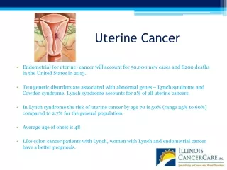
Introduction • The most common gynecologic cancer in developed countries. • In India, it ranks third amongst gynecologic cancer. • 70% cases are diagnosed in stage-I. • 5-year survival rate ~80%.
Risk factors Factors increasing risk • Increased exposure to unopposed estrogen • estrogen-replacement therapy, obesity, anovulatory cycles, estrogen-secreting tumors • Nulliparity • Years of menstruation • HNPCC family syndrome • Tamoxifen • Increasing age Factors decreasing risk • Grand multiparity • Oral contraceptives • Smoking • Physical activity
Role of Screening Routine screening NOT recommended. • Postmenopausal women on exogenous estrogens without progestins • Women from families with hereditary nonpolyposis colorectal cancer syndrome. • Premenopausal women with anovulatory cycles, such as those with polycystic ovarian disease
Patients in Whom a Diagnosis of Endometrial Cancer Should Be Excluded • All patients with postmenopausal bleeding • Postmenopausal women with a pyometra • Asymptomatic postmenopausal women with endometrial cells on a Pap smear, particularly if they are atypical • Perimenopausal patients with inter-menstrual bleeding or increasingly heavy periods • Premenopausal patients with abnormal uterine bleeding, particularly if there is a history of anovulation
Causes of Postmenopausal Bleeding • Atrophic endometritis/vaginitis • Endometrial or cervical polyps • Exogenous estrogens • Endometrial hyperplasia • Endometrial cancer • Miscellaneous (e.g., cervical cancer, uterine sarcoma, urethral caruncle, trauma)
Diagnosis & Pre-op investigations • Office endometrial biopsy: 10% false negative rate. • Fractional curettage under anesthesia: The gold standard. • Hysteroscopy : may identify polyps Fluid hysteroscopy may facilitate the abdominal dissemination of malignant cells, but there is no evidence that it has any impact on the disease-free survival. • X-ray chest, USG, CT scan, MRI, PET-CT, • Serum CA-125 • CBC, Renal function tests, Liver function tests, Blood sugar
Clinico-pathologic Types • Type-1 (low grade): ~90% Eo related- Arise on a background of hyperplasia. Long duration of unopposed estrogenic stimulation. Well-moderately differentiated. Favorable prognosis. • Type-2 (high grade): ~10% Eo non-related- Arise in atrophic endometrium. Non-estrogen dependent. Poorly differentiated or non-endometroid types. High risk of relapse and metastasis. Prognosis poor.
Ca-Endometrium & Ca-Ovary • ~8% Ca-endometrium associated with simultaneous presence of endometroid type Ca-ovary. • Synchronous: Independent primary • Both tumors are well differentiated and endometrial tumor is superficially invasive. • Metastatic: small, bilateral, or multi-nodular with surface implants and angiolymphatic invasion in the ovarian cortex. • D/Dx: by IHC and molecular genetic.
Spread Patterns • Direct extension to adjacent structures • Transtubal passage of exfoliated cells • Lymphatic dissemination • Hematogenous dissemination
Management of Endometrial Ca • 1900s Primary surgery • Mid 1930s Pre-operative RT • 1970s Primary surgery- clinical staging • 1988 FIGO Surgico-pathological staging • 2008 Revised FIGO staging
FIGO Surgico-pathologic staging (1988 vs 2008) St.-I Tumor confined to the corpus uteri IA No myometrial invasion IB Myometrial invasion < half IC Myometrial invasion ≥ half IA No or < half myometrial invasion IB Myometrial invasion ≥ half St.-II Cervical involvement IIA Cervical glandular involvement IIB Cervical stromal involvement II Tumor invades cervical stroma Revised FIGO staging. IJGO, 2009
FIGO Surgico-pathologic staging (1988 vs 2008) St.-III Local and/or regional spread IIIA Tumor invades the serosa of the uterus and/or adnexa and/or positive cytology IIIB Vaginal involvement parametrial involvement IIIC Pelvic and or para-aotic LN involvement IIIC1 Positive pelvic nodes IIIC2 Positive para-aortic nodes ± pelvic LNs St.-IV Tumor invades bladder and/or bowel mucosa or distant metastases IVA Invasion of bladder and/or bowel mucosa IVB Distant metastases
FIGO-Grade • Applies to Endometroid type; serous and clear cell carcinomas are considered to be high grade. • Grade 1: well formed glands with 5% solid, non-squamous areas. • Grade 2: 6%-50% solid non-squamous areas. • Grade 3:>50% solid non-squamous areas.
Steps of Surgical Staging • Peritoneal washings for cytology • Exploration of the abdomen & pelvis • Biopsy of any suspicious lesion • Total hysterectomy + BSO* • Pelvic & para-aortic lymphadenectomy * Ovarian preservation
Value of Surgical Staging Prognostic: • Accurately defines the extent of disease spread – Upstaging in up-to 23% clinical stage I • Determines the prognosis Adjuvant Rx: • Determines the need for and extent of adjuvant treatment Therapeutic:
Prognostic Value of Lymph Node Metastases -Morrow et al. Gynecol Oncol,1991
Morbidity of Lymphadenectomy Intra-operative: • Blood loss • Visceral injuries • Neuro-vascular injuries Post-operative: • Thrombo-embolism • Lymphocele • Lower limb/abdominal wall edema • GI complications
Controversies in the surgical staging • Complete surgical staging including pelvic and para-aortic lymphadenectomy for all patients. • Complete surgical staging NOT needed for any patient. Uterine risk factors are sufficient to identify high risk cases. • An identifiable group of intermediate/high risk patients will benefit from complete staging while those at low risk will not.
Predictors of LN Metastases • Depth of myometrial invasion • Tumor grade • Tumor size >2cm • Extra-uterine disease • Lymph vascular space invasion • Histologic sub-types – type II
Risk Stratification • Low risk:grade 1 or 2 histology with inner half of myometrial invasion,<2cm size, endometroid sub-type, without evidence of intra-peritoneal disease LN mets. 3-5% • High risk: grade 3 histology, any grade with deep myometrial invasion, non-endometroid sub-type or extra-uterine disease LN mets 10% - 20%
Extent of Lymphadenectomy Para-aortic LN Common iliac LN External iliac LN
Extent of Lymphadenectomy • Fifty percent of patients with pelvic node metastases will have additional para-aortic nodal metastases. • In 25% patients, para-aortic lymph node metastases can occur with negative pelvic nodes • Para-aortic LN involvement can occur above the IMA to the renal vessels directly
Role of Adjuvant Therapy Adjuvant therapy decisions -- Based on prognostic factors
Prognostic factors • Age • Histologic type • Histologic grade • Myometrial invasion • Vascular space invasion • Tumor size • Hormone receptor status • DNA ploidy and other biological markers
Mx of Advanced/recurrent disease • Multimodality Rx – Sx, RT, CT, HT. • Surgical cytoreduction in appropriately selected cases: Pts. with optimal cytoreduction have better survival than those with sub-optimal cytoreduction
Follow up protocol Every 3 mthly for 2 years Every 6 mthly for 3 years Annually life long History Clinical examination Vaginal cytology Radiological tests
Laparoscopy seems to be an appropriate alternative to open surgery Laparoscopic pelvic lymphadenectomy
Robotic Surgery Robotic surgery for Endometrial-Ca can be accomplished in heavier patients and results in shorter operating times and hospital stay, a lower transfusion rate, and less frequent conversion to laparotomy compared to laparoscopy. - Seamon et al. Gynecol Oncol,2009
Vaginal Surgery for Endometrial Ca • Vaginal hysterectomy with BSO: • Extreme obesity • Significant medical co-morbidity • Well differentiated tumors • Disadvantages • Upper abdominal exploration is not possible • Lymph nodes cannot be addressed
Endometrial Ca in Young Women Fertility preservation • Early stage, grade 1 and PR positive. • MRI to exclude significant myometrial invasion and adequate imaging of the ovaries • High dose progestins: megestrol acetate orally 160 to 320 mg/day or medroxyprogesterone acetate 200 to 500 mg/day • 40% of these patients will carry a successful pregnancy. • Hysterectomy is recommended once childbearing has been completed
Mx of Incompletely Staged Patient Review HPR- Grade, Invasion, adnexa + CT/MRI/ PET-scan No gross disease Gross disease IA,IB G1,2 IC, any grade IIA,IIB Surgical removal Observe Restaging Pelvic RT RT +/-CT
Conclusions • Disease of postmenopausal women. • Symptoms occur early in the course: most women have early stage disease at presentation. • Overall 5-year survival ~80%. • Type-1 (low grade, hormone sensitive): excellent prognosis • Type-2 (high grade, hormone independent): poor outcome.
Conclusions • Surgery is the primary modality; surgical staging offers the opportunity for the most accurate assessment of occult extra-uterine disease including nodal metastases. • Nodal metastasis is the most important risk factor. • The likelihood of nodal metastasis increases with the extent of disease and tumor grade. • Adjuvant Rx is needed in high risk cases.
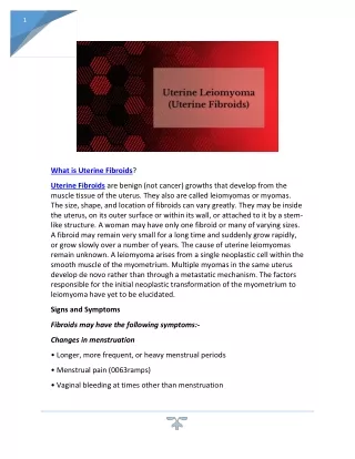
Uterine Sarcomas Pathological subtypes Incidence • Leiomyosarcoma25-30% • Endometrial stromal tumors 10-15% Endometrial stromal nodule Endometrial stromal sarcoma-low grade Undifferentiated sarcoma • Mixed epithelial-mesenchymal tumors Adenosarcoma 5% Carcinosarcoma (Mixed Mullerian Tumor) 45-50% Homologous Heterologous • Undifferentiated 5%
Management of uterine sarcomas • Surgery is the cornerstone of the treatment. • Total abdominal hysterectomy + B/L SO is the gold standard. • Debulking surgery in advanced cases. • ESS is hormone dependent so SO is indicated . • High recurrence rates even in early stage disease. • Adjuvant treatment has shown to significantly improvement in survival.