Download
1 / 19
200 likes | 970 Views
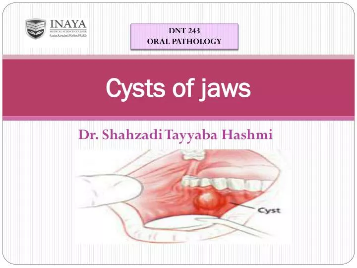
DNT 243 ORAL PATHOLOGY. Cysts of jaws. Dr. Shahzadi Tayyaba Hashmi. CYSTS OF JAWS. DEFINITION: Cysts are pathological fluid-filled cavities lined by epithelium

E N D
DNT 243 ORAL PATHOLOGY Cysts of jaws Dr. Shahzadi Tayyaba Hashmi
CYSTS OF JAWS DEFINITION: Cysts are pathological fluid-filled cavities lined by epithelium • Cysts are the most common cause of chronic swellings of the jaws. They are more common in jaws than in any other bone because of many rests of odontogenic epithelium remaining in the tissues.
INFLAMMATORY ODONTOGENIC CYSTS RADICULAR CYSTS • Residual • Lateral PARADENTAL CYSTS
Developmental Odontogenic cysts
ODONTOGENIC KERATOCYST(OKC) Pathology: • Odontogenic Keratocyst is derived from remnants of dental lamina Dental lamina: • The bands of epithelium that originates from oral epithelium and remain in the tissues after inducing tooth development
CLINICAL FEATURES OF ODONTOGENIC KERATOCYST • Peak incidence during second or third decade of life • Mandible is usually affected, primarily posterior body of mandible and ramus area • Unilocular or Multilocular • Multilocular OKC’s are consistent features of nevoid basal cell carcinoma syndrome( Gorlin Goltz Syndrome ) • Exhibits 25% to 60% of recurrence
RADIOGRAPHIC FEATURES • Odontogenic Keratocyst appear as well-definedradiolucent areas with a more or less rounded margins • Some are Unilocular, but majority are Multiloculated
HISTOPATHOLOGY Microscopic appearance has four major characteristics • A thin, uniform lining of ParakeratinizedSquamous epithelium, 6 to 10 cell thick • A palisaded layer of columnar or cuboidal basal cells • Corrugated layer of parakeratin 4. Lack of rete pegs
TREATMENT • Unilocular and small Multilocular cysts can be treated by Enucleation and bony cavity curetted • Large cysts have high rate of recurrence that’s why they need surgical resection and reconstruction with a bone graft
DENTIGEROUS CYST An Odontogeniccyst that surrounds the crownof an impacted tooth. It is usually derived from Reduced enamel Epithelium (residual Epitheliumthat surrounds the crown of tooth after enamel formation is complete)
CLINICAL FEATURES • Usually remain asymptomatic, but produce swelling or pain ,If it is large or INFLAMMED • More common in males as compared to females • Uncommon in children RADIOGRAPHIC APPEARANCE • Appear as well defined radiolucency surrounding the crown of an uneruptedtooth • In mandible, cyst may displace the associated tooth inferiorly into ascending ramus • In maxilla, it displaces associated tooth posteriorly
TREATMENT • Surgical ENUCLEATION • In case of a molar teeth, the associated tooth is usually extracted at the time when cyst is enucleated • In case of maxillary CUSPID tooth, cyst may be excised by MARSUPILIZATION (surgical curettage of cyst by creating a surgical window in cyst area)
ERUPTION CYST • An Odontogenic cyst with the histologic features of a Dentigerous cyst that surrounds a tooth’s crown that has erupted through bone but not soft tissue and is clinically visible as a soft fluctuant mass on the alveolar ridge
CLINICAL FEATURES AND TREATMENT CLINICAL FEATURES: • Affects children and involve teeth that have no predecessors (deciduous teeth) • Cyst lies superficially in gingiva overlying the unerupted tooth • Appears as a soft, rounded, bluish swelling MANAGEMENT: • Cyst roof may be removed to allow the tooth to erupt, but most eruption cysts burst spontaneously and require no treatment