Download
1 / 37
380 likes | 659 Views
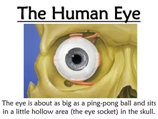
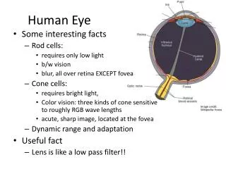
The Human Eye. 14.2 – Pages 449-455. A) Types of Vision . Monocular Binocular Peripheral Tunnel Vision. B) Layers of The Eye. Sclera (A) : Outer white part of the eye tough Resists punctures, is under fluid pressure from below Choroid (G) :

E N D
The Human Eye 14.2 – Pages 449-455
A) Types of Vision • Monocular • Binocular • Peripheral • Tunnel Vision
B) Layers of The Eye • Sclera (A): • Outer white part of the eye tough • Resists punctures, is under fluid pressure from below • Choroid (G): • Middle layer contains numerous blood vessels • Contains a black pigment darkens the interior of the eye • Ensures the eye ball is a “dark room” • Retina (L): • Contains the Photoreceptors Rods & Cones • Converts light into nerve impulse to the Occipital lobe
C) The Photoreceptors: Rods • Located in periphery • 120 million/Eye • Detects movement • Detects Black & White • Night Vision • Dim light (dawn & dusk) • Pigment: Rhodopsin Cones • Located in center • 6 million/Eye • Detects Color • Red, blue, green • Sharpness of vision • Bright Light (Day) • Pigment: Iodopsin Called “Visual Purple” and both contain Vitamin A
Colorblindness • Inherited through X Chomosome • The genetic information for certain cones in the eye are missing • Only lets the person see parts of the color spectrum clearly
Color Spectrum The first spectrum is normal and the next two are the spectrums that people with Protanopia and Deuteranopia see.
Test For Colorblindness • There is no cure for colorblindness at the present time. • There is a way to test for it.
Missing blue Missing green Missing Red
Fun facts…. • About 8% of men all men have some sort of color deficiency; about half of 1% of all women do • Humans are born color-blind. Cone cells don’t begin functioning until a baby is about 4 months old • Color-blindness is also known as Daltonism, named after John Dalton, who wrote the first scientific paper about the condition (which he had) in 1794. In 1995, 150 years after his death, reserarchersdeterminded that Dalton suffered from deuteranopia. How? They did a DNA analysis of his preserved eyeball.
Complete color-blindness, or rod monochromacy, is extrememly rare, except on the pacific island of Pohnpei, where 8% of the population has it. • Color-detecting cones work best in bright light. In very dim light only non-color detecting rods are used, which is why everything seems to be in black and white in dim light. • Rods are more numerous in the periphery of the retina. In dim light, use your peripheral vision – it sees better • Most mammals are dichromatic: they have two types of cone cells and can see fewer colors than we can. Honeybees, like humans, have three types. But honeybees can see colors in the ultraviolet range, humans can’t.
Source: Uncle John’s Slightly Irregular Bathroom Reader, 2004
D) Protection of the Eye: • Bony socket • Eyebrow deflects water and debris • Eyelashes have “blink” reflexes that respond to potential danger • Lacrimal Gland (tears): (top/side of eye) • Tears flow from top/temple to bottom/nasal • Tears contain an antibiotic • Tears drain into the nose
Protection cont’d: • Tarsal Gland (oil):(located in the lid) • Secretes an oil to moisturize the eye • Lids sweep the eye • Sclera to resist puncture
E) Eye Anatomy • Cornea (B)- outer transparent skin of the eye, no blood vessels, helps to focus image (refracts light towards pupil) • Iris (D)- circular muscle that helps control pupil size • Pupil (E)- allows light to enter the eye • Ciliary Muscles (F) – control the shape of the lens for focusing
Anatomy cont’d: • Lens (H) – focuses the image (inverts it too) • Aqueous Humour (I) – clear fluid in front between cornea and lens, circulates to nourish the cornea • Vitreous Humor (K) – clear pressurized fluid to maintain ball shape • Retina (L) – contains photoreceptors • Fovea Centralis (M) – 90% of cones are located here for color and sharpness of vision; most light sensitive area of the eye
Anatomy cont’d: • Blind Spot (N) – where optic nerve leaves the eye, no photoreceptors here • Optic Nerve (O) – contains 1 million axons to occipital lobe
F) Vision • Retina: Three layers of cells • Rods and cones are closest to choroids; generate nerve impulses in response to light (pigments change shape) • Middle layer contains bipolar cells • Innermost layer contain ganglionic cells, the axons of which form the optic nerve • Impulse travels from rods and cones bipolar cells ganglionic cells optic nerve optic chiasma occipital lobe
Vision cont’d… • Focusing: Bending Light • Light rays must be bent (refracted) to focus on the retina • Bent by: cornea, lens and to a lesser extent, humors • Accommodation Reflex – ability of lens to adjust in order to see close or distant objects. • For distant objects the lens is flat and the ciliary muscle relax • For close objects the lens becomes round and the ciliary muscles contract • With aging, lens loses some elasticity and is unable to accommodate. • Accommodation animation
Depth Perception • Depth perception is due to each eye forming an image from a slightly different angle. • Images on each side of the brain are interpreted as a whole.
G) Vision Defects: • Glaucoma: build-up of aqueous humor • Treatment: topical eye drops • Cause: high blood pressure; diabetes • Cataracts: clouding and/or hardening of lens • Treatment: removal; strong eyeglasses • Cause: age, trauma
Astigmatisms: irregular shaped cornea (or lens) • Treatment: glasses or contact lenses
Myopia:near sightedness; image is focused in front of fovea • Treatment: biconcave lens
Focusing Problems Animation • Hyperopia: far sightedness; image focused behind fovea • Treatment: biconvex lens
Eye Dissection • Eye Dissection – pg 463 in textbook • Identify the following parts: • Inside: • Lens • Humors (jelly) • Iris (scrape off cornea) • Retina (white, scrapes off easily) • Blind spot • Outside: • Optic Nerve (stump) • Cornea • Sclera
To Do: • Work on Human Eye Worksheet • Quiz tomorrow (Tuesday after the turkey coma!) on the EYE!