Download
1 / 60
3.82k likes | 9.02k Views
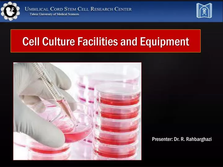
Cell Culture Facilities and Equipment. Presenter: Dr. R. Rahbarghazi. Small Tissue Culture Laboratory suggested for use by two or three persons. Medium-Sized Tissue Culture Laboratory suitable for five or six persons.

E N D
Cell Culture Facilities and Equipment Presenter: Dr. R. Rahbarghazi
Small Tissue Culture Laboratory suggested for use by two or three persons
Medium-Sized Tissue Culture Laboratory suitable for five or six persons
Tissue Culture Lab with Adjacent prep room with medium-sized tissue culture lab
Large Tissue Culture Laboratory; suitable for 20 to 30 persons
Laminar flow hood (Biological safety cabinet) • CO2 incubator (for most cells) • Inverted Microscope • Pipette aid • Aspiration pump • Centrifuge • Water bath • Cold storage • Cryopreservation Equipment
Sterilization equipment • Balances • Vortex • Water purification • pH meter • Magnetic stirrer • Micro pippettor • Cell counter • Video camera or CCD camera Additional or Optimal Equipment
1. Vertical mode 2. Horizontal mode Laminar flow hood (Biological safety cabinet)
Class I biosafety cabinet • Class II biosafety cabinet • Class III biosafety cabinet Vertical laminar flow hoods
Air is circulated away from operator • 100% of air is exhausted • Is optimal for radionuclides and volatile (toxic) materials • Dirty room air contaminates materials Class I Biosafety Cabinet
Type II-A • A front access opening with a carefully maintained inward flow • HEPA-filtered unidirectional airflow • HEPA-filtered exhaust air to the lab (30%) • 70% of the air re-circulated back into the laminar flow hood • Are not suitable for wok with radionuclides or volatile materials Class II Biosafety Cabinet
Type II B • Are conducted to exterior of building • Air are not re-circulated within the cabinets • Suitable for work with radionuclides and volatile materials
Providing highest level of protection for both material and operator • Is totally enclosed • Is under negative pressure • 100% of air is exhausted Class III Biosafety Cabinets
Inverted microscope Fluorescent inverted microscope Inverted Microscope Stereo or dissecting microscope
Vortex Water bath Balances
Flow cytometry integrates electronics, fluidics, computer, optics, software, and laser technologies in a single platform Flow cytometry
Advantages: • Provides individual measurements of cell fluorescence and light scattering • Enables us to individually sort or separate subpopulations of cells
FITC FITC FITC FITC Antibodies recognize specific molecules in the surface of some cells, especially stem cells Antibodies are artificially conjugated to fluorochromes (PE, FITC, RH, Alexa flour , … FITC Antibodies When the cells are analyzed by flow cytometry the cells expressing the marker for which the antibody is specific will manifest fluorescence. Cells who lack the marker will not manifest fluorescence FITC Fluorescence Activation Process (or Immunofluorescence) But not others
No reagents or probes required (Structural) Cell size (Forward Light Scatter) Cytoplasmic granularity (90 degree Light Scatter) Photsynthetic pigments Reagents are required 1. Structural DNA content DNA base ratios RNA content 2. Functional Surface and intracellular receptors. DNA synthesis DNA degradation (apoptosis) Cytoplasmic Ca++ Gene expression Intrinsic Extrinsic Cellular Parameters Measured by Flow
Immunofluorescence Cell Cycle Kinetics Cell Kinetics Genetics Molecular Biology Animal Husbandry (and Human as well) Microbiology Biological Oceanography Parasitology Bioterrorism Flow Cytometry Applications
FSC Correlates With Cell Size SSC Correlates With Internal Complexity
Fluorochromes Are Molecules That Emit Fluorescence Upon Excitation With Light • FITC (Fluorescein Isothiocyanate) • PE (Phycoerythrin) • PerCP (Peridinin Chlorophyll Protein) • APC (Allophycocyanin) • Some Fluorochromes Are Proteins, Some Are Small Organic Compounds • Ex. PE (Phycoerythrin)-Protein • Ex. FITC (Fluorescein Isothiocyanate)
Excitation Spectra Emission Spectra