Download
1 / 33
330 likes | 517 Views
Diagnostics. Gram Positive Rods. Classification. Characteristics of Spore Forming Rods. All are large Gram positive rods Make endospores Mainly found in soil, water and dust Highly diverse nutritional requirements Use simple and complex carbon sources

E N D
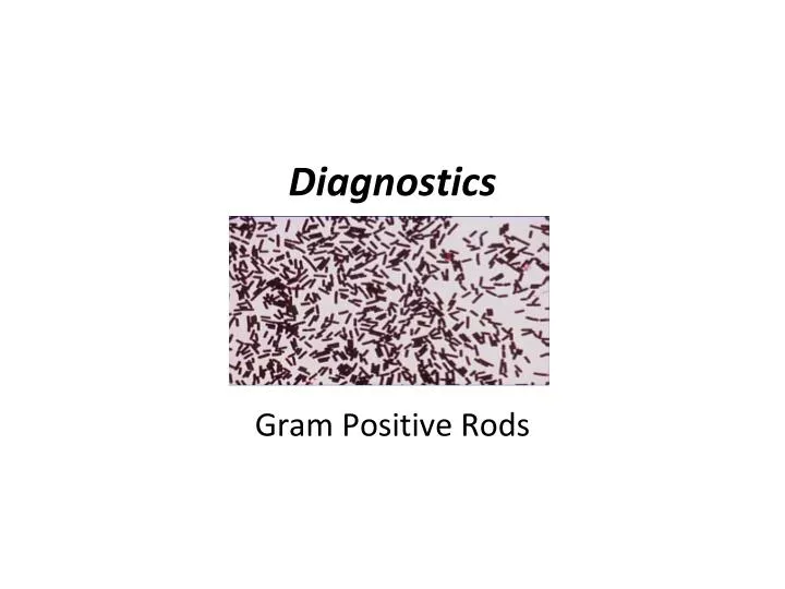
Diagnostics Gram Positive Rods
Characteristics of Spore Forming Rods • All are large Gram positive rods • Make endospores • Mainly found in soil, water and dust • Highly diverse nutritional requirements • Use simple and complex carbon sources • Their spores allow them to resist extreme conditions
Endospore Morphology • Location: • Terminal (a, d, e) • Subterminal (b) • Central (c, f). • Shape: • Circular (b, d) • Ellipsoid (a, c, e, f) • Spore diameter: • Non-deforming (a, b, c) • Deforming (d, e, f).
Medically Important Bacilli • Bacillus • Mostly harmless • A few opportunistic species • Bacillus cereus & Bacillus subtilis • Food poisoning • One pathogenic species • Bacillus anthracis • Anthrax
Medically Important Bacilli • Clostridium • Several pathogenic species • Clostridium perfringens • Gas gangrene • Clostridium tetani • Tetanus • Clostridium botulinum • Botulism • Clostridium difficile • Diarrhea Canned food that has not been sterilized properly; paralytic illness prolonged contraction of skeletal muscle fibers, neurotoxin produced by the bacteria
Identification: Metabolic Tests • Phenol red broth • Simple Carbon source: • Peptone (protein amino acids) • Desired sugar added • pH indicator • Phenol red • Yellow acid pH • Orange neutral pH • Red alkaline pH
Phenol Red Broths • Carbon utilization: • Sugar • Acid reaction (yellow) • or neutral (original) • Protein • Alkaline reaction (red)
Glucose Fermentation • Fermentation with acid accumulation: • Glucose pyruvate lactic and/or acetic acid + CO2 • Fermentation with accumulation of neutral products • Glucose pyruvate acetoin 2 butanediol + CO2
Methyl Red Test • Test for acid accumulation • Carbon Sources: Glucose and proteins • Indicator -methyl red; Added after growth • MR +: red (pH < 5.2) • MR - : Yellow (pH > 5.2) Neutral Acid
Voges-Proskauer Test Reagents VP: butanediol + -naphthol + KOH + O2 acetoin VP + = red VP - = Yellow - + Usual results of MR/VP: MR+/VP-; MR-/VP+ MR-/VP- Neutral Acid Acid produced No acetoin Neutral Acetoin
H2N C O + 2 H2O CO2 + H2O + 2 NH3 (NH4)2CO3 ammonium carbonate (alkaline) H2N Urea Amino acids Urea Utilization • Enzyme tested • Urease • pH Indicator • Phenol red (turns pink) Positive Negative
Complex Carbon Utilization • Too large to be transported inside • Requires exocellular enzymes for the external degradation into smaller units • Polysaccharides • Starch (amylase) • Lipids (lipase) • Tributyrin • Proteins (protease) • Casein (caseinase)
Before iodine addition After iodine addition Amylase – Starch Agar
2 H+ exterior Fp 2 e- interior 2 H+ 2 e- NADH + H+ FADH2 2 e- 2 H+ 3 H+ + 3 OH- 3 H2O 2 e- 3 H+ + 1/2 O2 Cyt o H+ H2O Q Fe-S Cyt b Aerobic RespirationElectron Transport Chain
Oxidase Test phenylenediamine • Cytochrome oxidase catalyzes the reduction of a final electron acceptor, oxygen • An artifcial e- donor, phenylenediamine, is used to reduce the cytochrome oxidase • If the enzyme is present, the colorless reagent (reduced state) will turn blue (oxidized state)
2H2O2 2H2O + O2 Product of respiration Damaging for DNA catalase Catalase Does bacteria make this? Detect bubbles. We add this. Add 3% H2O2 to bacterial growth bubbles (O2) Aerobic metabolism requires catalase
2 H+ Exterior Fp 2 H+ Interior 2 e- NADH + H+ FADH2 2 e- 2 H+ 3 H+ + 3 OH - 3 H2O 2 e- NO3- + 2 H+ (N = +5) nitrate Nitrate reductase Final e- acceptor 2 e- NO2- + H2O (N = +3) nitrite Q Fe-S Cyt b Anaerobic Respiration
NO3- + 2 H+ + 2 e- H2O + NO2- NO, N2O, NH2OH, NH3, N2 nitrite nitrate Nitrate is reduced to nitrite Nitrite is reduced No Nitrite Yellow Nitrate is reduced Production of Nitrite Red Nitrate is not reduced No Nitrite Yellow Nitrate Reductase Step 1: Test for nitrite NO2- + sulfanilic acid and alpha naphthylamine HNO2
NO3- + 2 H+ + 2 e- H2O + NO2- NO, N2O, NH2OH, NH3, N2 nitrite nitrate Nitrate is present Reduction to Nitrite Red Nitrate is absent Nitrite was reduced Yellow Nitrate Reductase (Cont’d) Step 2: Test for the presence of nitrate NO3- + Zn (s) NO2-
Diagnostics Gram Positive Cocci
Characteristics of Gram Positive Cocci • All are non sporulating • Mainly found amongst the natural flora of humans and animals • Fastidious (‘picky’) nutritional requirements • Use simple carbon sources
Cellular Aggregation of Gram Positive Cocci Micrococcus & Streptococcus Streptococcus Micrococcus Staphylococcus
Gram Positive Cocci of Medical Importance • Micrococcaceae • Staphylococcus aureus • Causes several types of infections, food infections and toxic shock (skin and respiratory tract) • Staphylococcus epidermidis • Cause opportunistic infections (catheters with biofilms) • Staphylococcus saprophyticus • Major cause of cystitis in women (bladder infection)
Gram Positive Cocci of Medical Importance • Streptococcaceae • Streptococcus pyogenes • Strep throat and flesh eating disease • Streptococcus agalactiae • Genital infections • Streptococcus mutans • Endocarditis • Streptococcus pneumonia • Otitis, meningitis, and pneumonia • Enterococcus spp. • Opportunistic infections
Identification: Metabolic Tests • Microccocus Vs Staphylococcus • Oxidase test • Micrococci are + • Staphylococci are – • Bacitracin (antibiotic) • Micrococci are sensitive • Staphylococci are resistant
Identification: Metabolic Tests • Differentitation of Staphylococcus species • Coagulase test • Coagulase positive staphylococci • S. aureus • Coagulase negative • All the other Staphylococci • Mannitol fermentation • S. aureus and some S. saprophyticus are positive • S. epidermidis is negative S. aureus S. epidermidis
Identification: Metabolic Tests • Differentiation of Streptoccocusspecies • Serological grouping according to Lancefield • Based on the type of carbohydrate in their cell wall • 8 groups (A-H and K-U) • Group A: β-hemolytic - Streptococcus pyogenes • Group B: β-hemolytic - S. agalactiae • Group C: α or γ hemolytic- S. viridans • Group D: γ –hemolytic – Enterococcus • Does not belong to any Lancfield group • α hemolytic- S. pneumoniae and S. mutans
Blood Hemolysis • Blood agar: • Discrimination according to hemolysis patterns • Alpha hemolysis – Incomplete hemolysis • Beta hemolysis- Complete hemolysis • Gamma hemolysis – No hemolysis
Identification: Metabolic Tests • Identification of Streptoccocus pneumoniae • Bile solubilization • Strep. pneumoniae is positive • Other Strep. are negative • Identification of Enterococcus • Bile-esculin test • Enterococcus is positive • Other Strep. are negative