Download
1 / 59
600 likes | 901 Views
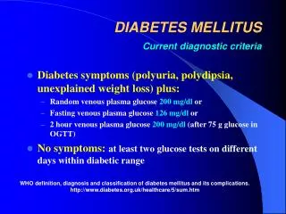
DIABETUS MELLITUS: ETIOLOGY, PATHOGENESIS, CLASSIFICATION, DIAGNOSTIC CRITERIA. Martynyuk L.P. Epidemiology. About 2 to 4 % of the world population is affected with DM The disease is more common: in persons after age 45 in obese individuals in certain ethnic groups

E N D
DIABETUS MELLITUS: ETIOLOGY, PATHOGENESIS, CLASSIFICATION, DIAGNOSTIC CRITERIA Martynyuk L.P.
Epidemiology • About 2 to 4 % of the world population is affected with DM • The disease is more common: • in persons after age 45 • in obese individuals • in certain ethnic groups • in those with a positive family history of DM • In patients with type 1 DM, complications from end stage renal disease are major cause of death, whereas patients with type 2 diabetes are more likely to have macrovascular diseases leading to myocardial infarction and stroke as main causes of the death
Diabetes mellitus (DM) - is endocrine – metabolic disease, which develops due to absolute or relative insulin insufficiency and characterized by chronic hyperglycemia, changes of different systems and organs of patient
The term DM • refers to the excretion of large quantities of sweet urine. Diabetes is an old word (from Greece “diabaino”) for siphon and means “dieresis”, mellitus (from Latina “mell”) means honey or “sweet” taste of a urine. Banting 1891 - 1941 Best 1899 - 1978
The clinical syndrome known as DM comprises a wide variety of symptoms, physical findings and laboratory abnormalities, in which multiple etiologic factors are involved, the pathophysiology is partly understood and treatment is unsatisfactory. • The hallmark of DM is hyperglycemia.
Insulin • Molecular weight of this peptic substance is 6000. • It consists of 51 aminoacidic parts from 16 differentaminoacids
Insulin • The most important biologic stimulator of insulin secretion is glucose
The action of insulin • Insulin is an anabolic hormone (promotes the synthesis of carbohydrates, proteins, lipids and nucleic acids). The most important target organs for insulin action are: • the liver, muscles and adipocytes. • The brain (nervous tissue), retina, lens and blood cells are unresponsive to insulin. • It has no direct influence on kidney also
The effects of insulin oncarbohydrate metabolism • stimulation of glucose transport across muscle and adipose cell membranes; • regulation of hepatic glycogen synthesis; • inhibition of glucose formation – from glycogen (glycogenolysis) and – from amino-acid precursors (glyconeogenesis). • The result of these actions is a reduction in blood glucose concentration.
Protein metabolism: • the transfer of amino acids across plasma membranes; • stimulation of protein synthesis; • inhibition of proteolysis.
Lipid metabolism: • incorporation of fatty acids from circulating triglyceride into adipose triglyceride; • stimulation of lipid synthesis; • inhibition of lipolisis.
Nucleic acids metabolism: • stimulation of nucleic acid synthesis by stimulating the formation of adenosine triphosphate (ATP), DNA and RNF. Other effects: • stimulation of the intracellular flew of potassium, phosphate and magnesium in the heart; • inhibition of inotropic and chronoropic action (unrelated to hypoglycemia).
Absolute 1. Genetic disorders 2. Autoimmune damaging of β-cells 3. Damaged caused by virusessuch as mumps, or Coxsackie B4 4. Toxic influence on β-cells 5. Diseases of pancreatic gland Relative β-cells Insulin transport Receptors (tissue insensitivity) Insulin insufficiency
Etiologic classification of DM (1999) I. Type 1 of DM (destruction of β-cells which mostly leads to absolute insulin insufficiency): • autoimmune; • idiopathic. II. Type 2 of DM (resistance to insulin and relative insulin insufficiency or defect of insulin secretion with or without resistance to insulin). III. Other specific types: • genetic defects of β-cells function; • genetic defects of insulin action; • pancreatic diseases (chronic pancreatitis; trauma, pancreatectomy; tumor of pancreatic gland; fibrocalculosis; hemochromatosis); • endocrine disease (acromegaly, thyrotoxicosis, Cushing’s syndrome); • drug exposures ; • infections and others. IV. Gestation diabetes.
Stages of DM development • I. Prediabetes (risk factors or predispose factors). • II. Impaired glucose tolerance (latent DM). • III. Clinical manifestation of DM.
Prediabetes (risk factors or predispose factors) • obesity; • positive family history of DM; • persons which were born with weight more than 4,0 kg; • women who had =children with weight more than 4,0 kg; =abortions and =dead child in anamnesis; • persons with: = atherosclerosis, hypertension; = auto-immune diseases; = furunculosis; = rubella, mumps, Coxsackie virus, infectious hepatitis, cytomegalovirus, infection mononucleosis; • endocrine disorders
Stages of compensation • Criteria of compensative stage.1.Patient hasn’t new complains.2.Fast serum glucose level is normal.3.Glucose in urine is absent.4.Glucose level fluctuation is under 4.4-5.5 mmol/l during the day .5. HbA1c is 6,0 – 7,0 for the 1 type of DM, 6,0-6,5 for the 2 type of DM 6.Comatose and precomatose status are absent.
Criteria of decompensative stage: • 1. Postprandial glycemia is >9,0 mmol/l. • 2. HbA1c is higher then 7,5 for the 1 type of DM, 7,0 for the 2 type of DM • 3. Comatose or precomatose status are present.
Duration of DM 1.Stabile - glucose level fluctuation is under 4.4-5.5 mmol/l during the day - comatose or precomatose status are absent. 2.Labile glucose level fluctuation is over 4.4-5.5 mmol/l during the day - or comatose and precomatose status are present.
Type I, or insulin-dependent diabetes mellitus (IDDM) • is characterized by pancreatic islet beta cell destruction and absolute insulinopenia. • This individuals are ketosis prone under basal conditions. The onset of the disease is generally in youth, but it can occur at any age. Patients have dependence on daily insulin administration for survival.
Pathogenesis of type I DMincludes the following: I. Current formulation of the genetic predisposition, conferred by diabetogenic genes on the short arm of chromosome 6, either as part of it or in close proximity to the major histocompatibility complex (MMHC) region (more than 95 % of type I diabetes individuals are HLA DR3, DR4 or DR3/DR4; on the other hand, HLA DR2 confers protection against the development of type I DM); II. Putative environmental triggers (possibly viral infections (Coxsackie B, rubella) or chemical toxins (nitrosourea compounds)) that in genetically susceptible individuals might play a role in initiating the disease process. III. An immune mechanism gone awry, either initiation of immune destruction or loss of tolerance, IV. leading to slow, progressive loss of pancreatic islet β-cells (50%) and V. eventual clinical onset of type I diabetes. VI.Total destruction of β-cells
Type II, or noninsulin-dependent diabetes mellitus (NIDDM) • Type 2 is the most common form of diabetes, accounting for 95 – 90 % of the diabetic population. Most investigators agree that genetic factors underlie Type 2 DM, but it is probably not caused by defects at a single gene locus. - Obesity, - diet, - physical activity, - intrauterine environment, - stress are among the most important environmental factors which play a role in the development of the disease.
Pathogenetic and clinical difference oftype I and type II DM
Pathogenetic and clinical difference oftype I and type II DM
Pathogenetic and clinical difference oftype I and type II DM
Clinical presentation The classic manifestation of type 1 DM include: • polyurea ( when the level of the blood glucose is more then 9 mmol/l, glucosurea arises). • polidipsia (as more water is excreted, the body requires more water intake); • polyphagia (as a result of lack of energy); • loss of weight (energy (calories) is lost as glucose in the urine. Loss of water itself also contributes to weight loss. Increased proteolysis with mobilization of aminoacids leads to progression of protein catabolism and loss of weight, mostly in muscle mass); • fatigue and weakness (probably occur as a result of decre-ased glucose utilization and electrolyte abnormalities); • acidosis (develops due to increased lipolysis which cause the release of free fatty acids, which are metabolized to ketones by the liver)
Presenting signs and symptoms of type 2 DM include: • polyurea, • polydipsia, • polyphagia; but they are not prominent The majority of patients (80 – 85 %) are obese, but it can also occur in thin persons.
Classification of chronic (long-term) complications of DM. I. Diabetic angiopathy: 1. Microangiopathy: 1) nephropathy; 2) retinopathy; 3) angiopathy of lower extremitas. 2. Macroangiopathy: 1) heart (ischemic heart disease); 2) brain 3) angiopathy of lower extremities. II. Diabetic neuropathy: 1) central (encephalopathy); 2) peripheral; 3) visceral (dysfunction of inner organs).
The long-term degenerative changes in the blood, vessels, the heart, the kidneys, the nervous system, and the eyes as responsible for the most of the morbidity and mortality of DM.
Skin • The skin is dry and itch • Infections of the skin by bacteria and fungi, candidiasis of the external female genitalia, hyperkeratosis, nail disorders are common in the patients with DM but nothing is specific with regard to their development.
The most common skin lesion is diabetic dermopathy (it is characterized by brown, atrophic, well-demarcated areas in the pretibial region which resemble sears), besides patients sometimes have xanthoma diabeticorum, which is usually located on the buttocks, elbows and knees, look like eruptions (but is not really diabeticorum since it occurs in the patients with lipoprotein abnormalities, particularly hyperchylomicronemia, whether or not patient has DM)
Subcutaneous adipose tissue • The abdomen type of obesity is common in patients with type II DM. • Sometimes generalized subcutaneous adipose tissue atrophy can be observed in diabetics.
Bones and joints • Osteoporosis and osteoarthropaphy can be find in patients with DM also. • Diabetic chairopathy (decreasing of the movements of joints)
Gastrointestinal tract • Paradontosis, gastritis with decreased secretion ability, gastroduodenitis, hepatosis are common in patients with DM. • Visceral dysfunction gastrointestinal tract: esophageal neuropathy (disturbances of peristalsis in the body of the esophagus.) diabetic gastroparesis (irregular food absorption and is characterized by nausea, vomiting, early satiety, bloating and abdomen pain.); diabeticenteropathy (diarrhea (mostly at night time, postural diarrhea), constipation, malabsorption and fecal incontinence)
Cardiovascular system (CVS). • Diabetic autonomiccardiopathy: - orthostatic hypotension (is characterized by dizziness, vertigo, faintness, and syncope upon assumption of the upright posture and is caused by failure of peripheral arteriolar constriction.); - tachicardia (but it does not occur in response to hypotension because of sympathetic involvement). • Dismetabolic cardiomyopathy (IHD, rhythm disturbances)
Ischemic heart disease • Cardiovascular changes tend to occur earlier in patients with DM when compared with individuals of the same age. • Frequency of myocardial infarction (MI) and mortality is higher in diabetics than that in nondiabetis of the same age. • The prognosis is even worse if ketoacidosis, or other complications of DM are present. 4. Diabetic patients have more complications of MI (arrhythmias, cardiogenic shock and others) than nondiabetic ones. 5. Often can observe atypical forms (without pain). 6. Male : female = 1 : 1 (nondiabetics = 10 : 1).
Respiratory system • Mucomycosis of the nasopharinx, sinusitis, bronchitis, pneumonia (prolonged duration, slow reccurency), tuberculosis are more common in patients with diabetes than in nondiabetics.
Kidneys and urinary tract. • Inflammation processes (10 – 30 %): pyelitis, pyelonephritis, cystitis • Diabetic cystopathy or neurogenic vesicle dysfunction (enlargement of the volume of the cyst bladder, insidious onset and progression of bladder paralysis with urinary retention, decreasing of quantity of urinations) • Diabetic nephropathy Sexual disorders: • retrograde ejaculation (which is caused by dysfunction of the pelvic autonomic nervous system); • impotence, and sometimes decreased libido;
Diabetic nephropathy(by Mogensen) I. Hyperfunction of kidneys - increased renal blood circulation; increased glomerular filtration rate (GFR) (> 140 ml/min); hypertrophy of kidneys; normoalbuminuria (<30 mg/day). II. Stage of initial changes of kidney structure. mesangial changes due to accumulation of immunoglobulins (IgG, IgM), complement and other nonimmunologic proteins (lipoproteins, fibrin); high GFR; - normoalbuminuria III. Initial nephropathy. - microalbuminuria (30 to 300 mg/day); - high or normal GFR; - periods of blood hypertension
I IV. Nephropathy or nephrotic stage. • - persistent proteinurea (>500 mg/day); • - normal or decreased GFR; • - persistent blood hypertension. V. Chronic renal failure or uremia. - decreased GFR; - blood hypertension; - increased serum creatinine - signs of intoxication.
Eyes • Ceratities, retinatis, chorioretinatis, cataracts, glaucoma • Diabetic retinopathy - Evidence of retinopathy, rarely present at diagnosis in type I DM, is present in up to 20 % of type II DM patients at diagnosis. About 85 % of all diabetics eventually develop some degree of retinopathy - the initial retinal changes (seen on the ophthalmoloscopic examination) does not significantly alter vision, but it can lead to processes that cause blindness
Diabetic retinopathy (is classified according to the changes seen at background during ophthalmoscopic examination) I stage. Background retinopathy II stage. Maculopathy or preproliferative retinopathy III stage. Proliferative retinopathy
Diabetic retinopathy I stage. Background retinopathyis usually the earliest sigh and consists of retinal microaneurysms, hard and soft exudates.
Diabetic retinopathy II stage. Maculopathy or preproliferative retinopathyis characterized by macular edema and/or hemorrhages. III stage. The hallmark of proliferative retinopathyis neovascularization, i.e., growth of new vessels in areas of hypoperfusion. Adhesion of the vessels to the vitreous leads to retinal detachment, vitreous hemorrhage and others. The prognosis is extremely poor. 5 years after recognition of this complication 50 % of the patients are blind.