Download
1 / 42
420 likes | 504 Views
Learn modern diagnostic processes for treating patients with musculoskeletal lesions in the maxillofacial area. Explore techniques to assess joints, muscles, and explore dental assistant duties.

E N D
Diagnosis and treatment of patients with lesions of the musculoskeletal system maxillo-facial area. Modern diagnostic process in prosthetic dentistry.
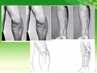
The joints are palpated as the patient opens and closes to detect signs of dysfunction. The masseter muscle can be palpated extraorally by placing your fingers over the lateral surfaces of the ramus of the mandible.
Fingers are placed over the patient's temples to feel the temporalis muscle.
The little finger is inserted facial to the maxillary teeth and around distal to the pterygomaxillary, or hamular, notch to palpate the lateral pterygoid muscle. The index finger is used to touch the medial pterygoid muscle on the inner surface of the ramus.
The sternocleidomastoid muscle is grasped between the thumb and forefingers on the side of the neck. The muscle can be accentuated by a slight turn of the patient's head. The trapezius muscle is felt at the base of the skull, high on the neck.
The distance between maxillary and mandibular incisors is measured when the patient is instructed to open "all the way" (A). If the patient can only open part way (B), the cause should be determined.
If opening is limited, the patient should be instructed to use a finger to indicate the area that hurts.
Rubber gloves, a surgical mask, and eye protection are important for safeguarding dental office personnel.
Auricular palpation of the posterior aspects of the temporomandibular joints.
Maximum opening of more than 50 mm (A) and lateral movement of about 1 2 mm (B) are normal.
Muscle palpation. A, The masseter. B, The temporal muscle. C, The trapezius muscle. D, The sternocleidomastoid muscle. E, The floor of the mouth.
Palpation is best done bilaterally, simultaneously asking the patient to identify any differences between left and right.
Smile analysis is an important part of the examination, particularly when anterior crowns or fixed dental prostheses are being considered. A, Some individuals show considerable gingival tissue during an exaggerated smile. B, Others may not show the gingival margins of even the central incisors.
The "negative space" between the maxillary and mandibular teeth is assessed during the examination.