Download
1 / 54
540 likes | 942 Views
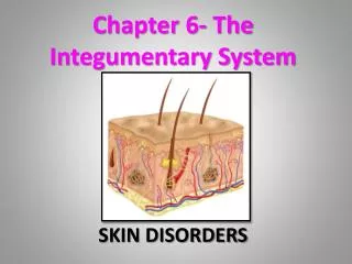
Chapter 3 INTEGUMENTARY SYSTEM. The skin is the largest and most visible organ of the body Weighs 8-10 pounds and covers an area of 22 square feet Combining forms: Derm/o Cutane/o Dermatology is the medical specialty.

E N D
Chapter 3INTEGUMENTARY SYSTEM • The skin is the largest and most visible organ of the body • Weighs 8-10 pounds and covers an area of 22 square feet • Combining forms: • Derm/o • Cutane/o • Dermatology is the medical specialty
The skin (body covering system) is a readily visible reflection of one’s health
FUNCTIONS OF THE SKIN (5) • (1) Protection • Body’s first level of defense against invasion by bacteria and other harmful agent • Protects against mechanical injury • Guards against excessive exposure to sun • (2) Regulation (temperature control) • Raises or lowers body temperature as necessary
FUNCTIONS (continued) • (3) Sensation • Contains millions of microscopic nerve endings • Act as sensory receptors for pain, touch, heat, cold, pressure, and pleasure
FUNCTIONS (Continued) • (4) Secretion • Glands secrete: • Perspiration (sweat glands) • Oils – sebaceous Glands
FUNCTIONS • (5) Water retention • Acts as a barrier between internal organs and the environment
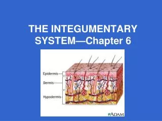
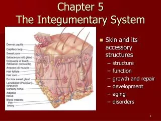
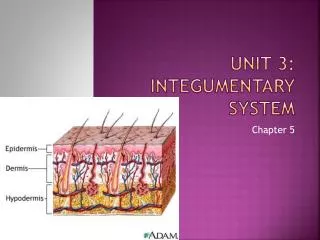
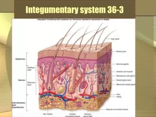
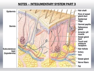
SKIN ANATOMY • Made up of 3 layers: • Epidermis – outer (top) layer (epi) • Dermis - inner support layer (middle) • Subcutaneous – bottom layer –adipose tissue (sub)
EPIDERMIS LAYER • Consists of 5 layers • New skin cells are formed in the lowest level • New skin cells contain keratin • Melan/o – brown/black color • Melanocytes are cells that produce melanin (pigment) which give skin and hair its color. • New skin cells move up to the top layer-all replaced about every 28 days.
DERMIS LAYER • Composed of dense, fibrous connective tissue called collagen. • Contains nerves, sensory receptors , blood vessels. • Essential to thermoregulation • Contains hair follicles • Contains sebaceous and sweat glands
SEBACEOUS GLANDS • Sebum – oily substance made up of fats and lipids • protects skin
SWEAT GLANDS • Glands all over the body • Duct of gland • Conduit to surface for sweat • Sweat cools body as the moisture evaporates • Palms of hands • Feet
SUBCUTANEOUS LAYER • Fat cells stored for energy • Provides heat insulation • Connects the skin to underlying structures
HAIR • Formed within follicles • Papilla at base of follicle • Shaft is visible part • Hair fiber is filled with keratin • Melanocytes at root of follicles • Pigment provides color • Gray hair- no melanin produced
Nails • Composed of hard keratin
PHYSIOLOGY OF THE SKIN • Identification function • Unique facial characteristics, hair, skin color, fingerprints • Communication • Expressions – expressing our emotions • Sensory functions • Pain, pressure, temperature • Production of vitamin D • Exposure of skin to ultraviolet light
PHYSIOLOGY (con ‘t) • Protection • Skin is first line of defense against the outside world – goal is integrity • Skin is waterproof and tough • Melanin prevents harmful ultraviolet rays from penetrating • Keratin is a waterproofing agent • Stratum corneum (outermost, horny cell layer) protects against pathogens, chemicals, prevents tears
TEMPERATURE CONTROL • Raises and lowers body temperature as necessary • When body needs to lose heat: • Blood vessels in skin dilate – bringing blood to surface for cooling by radiation • Sweat glands secrete more sweat for cooling by evaporation
TEMPERATURE CONTROL(con’t) • When body needs to conserve heat, nervous system causes constriction of skin’s blood vessels • Allows more heat-carrying blood to circulate to the muscles and other vital organs • Skeletal Muscles • Produce heat during movement • Raises body’s core temperature • Produce heat during movement • Increases body’s core temperature
WELLNESS & ILLNESS • General assessment • Overview • General hygiene • Well developed, well nourished, or other ? • Inspection and palpation • Color, appearance, temperature, turgor • Vascularity • Lesions or abnormalities
INFANTS • Commonly seen conditions • Vernix caseosa • Lanugo – fine hair • Mongolian spots • Café au lait spots • Port-wine stain (nevus flammeus) • Erythema toxicum-rash • Acrocyanosis • Physiologic jaundice
ADOLESCENTS • Acne • Active sabaceous glands • Types: • Open comedones (blackheads) • Closed comedones (whiteheads) • May also include • Papules • Pustules • Nodules
PREGNANT WOMEN • Skin stretches: • Striae –stretch marks • Caused by elastic collagen fibers breaking down • Linea nigra – midline of abdomen • Chloamsa – hyperpigmentation • Vascular spiders – tiny red lines
ADULTS & SENIORS • Middle-aged adults • Skin changes • Angiomas on trunk • Skin tags • Increased/decreased hair growth • Alopecia – male pattern balding • Seniors • Skin’s gradual breakdown: dryness,wrinkling • Skin more fragile • Trauma – senile purpura • Liver spots • Keratoses
GENERAL SKIN CONDITIONS • Ringworm –fungus • Dermatitis –inflammation associated with an allergic reaction • Allergen touches skin – contact dermatitis • Ingestion of allergic substance • Rash may be urticarial – hives, itchy • Atopic dermatitis • Chronic inflammation – usually caused by allergy • Psoriasis • Seborrhea – cradle cap, dandruff • Lesions • Nevi – birth marks/beauty marks • Hemangioma – collection of blood vessels
SKIN CANCERS • Melanoma • Cancer that develops in the pigment cells • An overgrowth of melanocytes • Most dangerous form of skin cancer • Can spread ( metastasize) • A-B-C-D changes in lesion
MELANOMA • A-B-C-D changes that indicate malignancy • Asymmetry • Border • Color • Diameter
SKIN CANCERS (Continued) • Cancers of the epithelial cells • Appear in areas exposed to sunlight, such as the face • Basal cell • 75% of a skin cancers • Smooth, pearly papule • Slow growing • Do NOT metastasize • Treatment: surgical removal
SKIN CANCERS (Continued) • Squamous cell • Painless, firm red nodule or plaque • May develop surface scales, ulcerations or crusting • Tends NOT to metastasize • Treatment: surgical removal • Kaposi sarcoma • Associated with AIDS • Distinct brown lesions on the legs • Can metastasize
Primary Skin Lesions • Skin signs are objective evidence of an illness or disorder • Various lesions are described in specific terms • Primary or initial lesions • Secondary – result of primary lesions
Cyst • An encapsulated, fluid-filled area in the dermis or subcutaneous layer
Bulla • A fluid-filled lesion larger than 1 cm that is thin-walled and ruptures easily • Example – blister
Macule • A round, flat, pigmented area • Example − measles, freckles
Nodule • A solid, raised area larger than 1 cm • A larger papule • Example − acne vulgaris
Papule • A palpable (something that can be felt) lesion that is rounded, solid, and raised (less than 1 cm) • Example – pimple, mole
Plaque • An area of papules that are merged to form a lesion larger than 1 cm • Example − psoriasis
Pustule • A round, raised, pus-filled lesion • Example – chicken pox in acute stage
Urticaria • An intensely itchy (pruritic) area of wheals that have merged • Wheals are raised, red, and irregularly shaped • Due to an allergic reaction • Usually itchy
Vesicle • A raised, clear fluid-filled sac,up to 1 cm in size • Example – blister
SECONDARY SKIN LESIONS • Primary skin lesions that have changed as a result of manipulation (scratching) or natural/pathologic progression
Crust • A thickened, dried area from broken pustules or vesicles • Examples: impetigo, chicken pox (when drying) scabs, eczema
Erosion • A superficial, scooped-out area that does not extend into the dermal layer
Excoriation • Reddened abrasions, usually from scratching • Example – scabies, insect bites
Fissure • A linear crack or slit that extends through the epidermis into the dermis • Example – athlete’s foot
Keloid • Excess scar tissue; most commonly seen in African-Americans • Forms at site of injury or incision
Scale • A thin flaky, dry, silvery or white form of shedding keratin cells • Examples – ringworm, eczema, psoriasis
Scar • Connective tissue that remains after a skin lesion has healed • Examples − chicken pox scar, acne
Ulcer • A deep depression that extends into the dermis • Also called erosion,crater • Pressure ulcer (decubitus or bedsore) • Caused by lack of circulation to avulnerable area – a bony prominence • Can lead to necrosis • Potentially very serious
HAIR & NAILS −GENERAL CONDITIONS • Hair • Hirsutism − abnormal hair growth (increased) • Alopecia – absence of hair, baldness • Nails • Paronychia – inflammation around nail • Onycholysis – loosening of nail plate
DIAGNOSING & TREATING: TESTS • Biopsy • To determine diagnosis • Patch test • To identify specific allergies • Scratch test • Scratching surface of skin with allergens • Intradermal injections • Local inflammatory response • PPD – TB • Wood’s lamp • Ultraviolet light to diagnose tinea (ringworm)
PROCEDURES • Surgical • Debriding – burns (eschar) • Laser – to remove lesions, unwanted hair • Dermabrasion • Removes scars • Chemical peel • Removes small lines and scars • Cosmetic: • Fillers and injectables – Botox • Topicals • Surgery • Many types of plastic surgery